{"title":"喹司他酯体内外抗刚地弓形虫活性研究。","authors":"Hui-Jie Qiu, Wen-Bin Zheng, Ting Zeng, Shu-Feng Yang, Dai-Ang Liu, Li-Yan Wang, Zhi-Rong Liu, Xing-Quan Zhu, Chun-Xue Zhou","doi":"10.1128/aac.01819-24","DOIUrl":null,"url":null,"abstract":"<p><p><i>Toxoplasma gondii,</i> an opportunistic pathogen, poses severe threats to immunocompromised individuals and fetuses of newly infected pregnant women. The current gold-standard treatment, a combination of pyrimethamine and sulfadiazine, is limited by severe adverse events, necessitating the development of novel therapeutic agents. <i>In vitro</i>, a CCK8 assay demonstrated that quisinostat inhibited HeLa cell proliferation in a dose-dependent manner, with a CC<sub>50</sub> of 8.22 nM. Regarding <i>T. gondii</i> tachyzoites, quisinostat exhibited time-dependent inhibition of extracellular parasite activity and suppressed intracellular parasite proliferation, with an EC<sub>50</sub> of 25.84 pM, and a high selectivity index (SI = 318.11). Quisinostat disrupted the <i>T. gondii</i> lytic cycle by decreasing invasion rates, inducing G1 cell cycle arrest, reducing replication, and shrinking plaque size. Ultrastructural analysis indicated that quisinostat treatment led to membrane damage, enhanced lactate dehydrogenase (LDH) release, and apoptotic cell death in tachyzoites, whereas no significant change in reactive oxygen species (ROS) levels was detected. Proteome analysis identified 77 upregulated and 205 downregulated proteins, which were enriched in functions associated with protein dephosphorylation and ion transport, as well as pathways, such as non-homologous end-joining. Molecular docking studies revealed a strong interaction between quisinostat and <i>T. gondii</i> HDAC3. <i>In vivo</i>, treatment with quisinostat increased the survival time of mice infected with virulent RH strain. In mice infected with low-virulent ME49 tachyzoites, quisinostat treatment decreased parasite burden in multiple organs and increased the survival to 80%. Taken together, these findings demonstrate that quisinostat has potent anti-<i>Toxoplasma</i> activity both <i>in vitro</i> and <i>in vivo</i>, which offers promise for treatment of human toxoplasmosis.</p>","PeriodicalId":8152,"journal":{"name":"Antimicrobial Agents and Chemotherapy","volume":" ","pages":"e0181924"},"PeriodicalIF":4.5000,"publicationDate":"2025-09-03","publicationTypes":"Journal Article","fieldsOfStudy":null,"isOpenAccess":false,"openAccessPdf":"https://www.ncbi.nlm.nih.gov/pmc/articles/PMC12406659/pdf/","citationCount":"0","resultStr":"{\"title\":\"<i>In vitro</i> and <i>in vivo</i> activity of quisinostat against <i>Toxoplasma gondii</i>.\",\"authors\":\"Hui-Jie Qiu, Wen-Bin Zheng, Ting Zeng, Shu-Feng Yang, Dai-Ang Liu, Li-Yan Wang, Zhi-Rong Liu, Xing-Quan Zhu, Chun-Xue Zhou\",\"doi\":\"10.1128/aac.01819-24\",\"DOIUrl\":null,\"url\":null,\"abstract\":\"<p><p><i>Toxoplasma gondii,</i> an opportunistic pathogen, poses severe threats to immunocompromised individuals and fetuses of newly infected pregnant women. The current gold-standard treatment, a combination of pyrimethamine and sulfadiazine, is limited by severe adverse events, necessitating the development of novel therapeutic agents. <i>In vitro</i>, a CCK8 assay demonstrated that quisinostat inhibited HeLa cell proliferation in a dose-dependent manner, with a CC<sub>50</sub> of 8.22 nM. Regarding <i>T. gondii</i> tachyzoites, quisinostat exhibited time-dependent inhibition of extracellular parasite activity and suppressed intracellular parasite proliferation, with an EC<sub>50</sub> of 25.84 pM, and a high selectivity index (SI = 318.11). Quisinostat disrupted the <i>T. gondii</i> lytic cycle by decreasing invasion rates, inducing G1 cell cycle arrest, reducing replication, and shrinking plaque size. Ultrastructural analysis indicated that quisinostat treatment led to membrane damage, enhanced lactate dehydrogenase (LDH) release, and apoptotic cell death in tachyzoites, whereas no significant change in reactive oxygen species (ROS) levels was detected. Proteome analysis identified 77 upregulated and 205 downregulated proteins, which were enriched in functions associated with protein dephosphorylation and ion transport, as well as pathways, such as non-homologous end-joining. Molecular docking studies revealed a strong interaction between quisinostat and <i>T. gondii</i> HDAC3. <i>In vivo</i>, treatment with quisinostat increased the survival time of mice infected with virulent RH strain. In mice infected with low-virulent ME49 tachyzoites, quisinostat treatment decreased parasite burden in multiple organs and increased the survival to 80%. Taken together, these findings demonstrate that quisinostat has potent anti-<i>Toxoplasma</i> activity both <i>in vitro</i> and <i>in vivo</i>, which offers promise for treatment of human toxoplasmosis.</p>\",\"PeriodicalId\":8152,\"journal\":{\"name\":\"Antimicrobial Agents and Chemotherapy\",\"volume\":\" \",\"pages\":\"e0181924\"},\"PeriodicalIF\":4.5000,\"publicationDate\":\"2025-09-03\",\"publicationTypes\":\"Journal Article\",\"fieldsOfStudy\":null,\"isOpenAccess\":false,\"openAccessPdf\":\"https://www.ncbi.nlm.nih.gov/pmc/articles/PMC12406659/pdf/\",\"citationCount\":\"0\",\"resultStr\":null,\"platform\":\"Semanticscholar\",\"paperid\":null,\"PeriodicalName\":\"Antimicrobial Agents and Chemotherapy\",\"FirstCategoryId\":\"3\",\"ListUrlMain\":\"https://doi.org/10.1128/aac.01819-24\",\"RegionNum\":2,\"RegionCategory\":\"医学\",\"ArticlePicture\":[],\"TitleCN\":null,\"AbstractTextCN\":null,\"PMCID\":null,\"EPubDate\":\"2025/7/31 0:00:00\",\"PubModel\":\"Epub\",\"JCR\":\"Q2\",\"JCRName\":\"MICROBIOLOGY\",\"Score\":null,\"Total\":0}","platform":"Semanticscholar","paperid":null,"PeriodicalName":"Antimicrobial Agents and Chemotherapy","FirstCategoryId":"3","ListUrlMain":"https://doi.org/10.1128/aac.01819-24","RegionNum":2,"RegionCategory":"医学","ArticlePicture":[],"TitleCN":null,"AbstractTextCN":null,"PMCID":null,"EPubDate":"2025/7/31 0:00:00","PubModel":"Epub","JCR":"Q2","JCRName":"MICROBIOLOGY","Score":null,"Total":0}
In vitro and in vivo activity of quisinostat against Toxoplasma gondii.
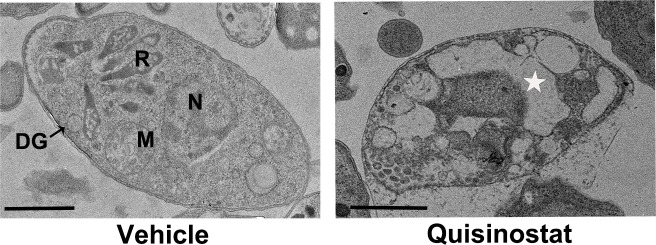
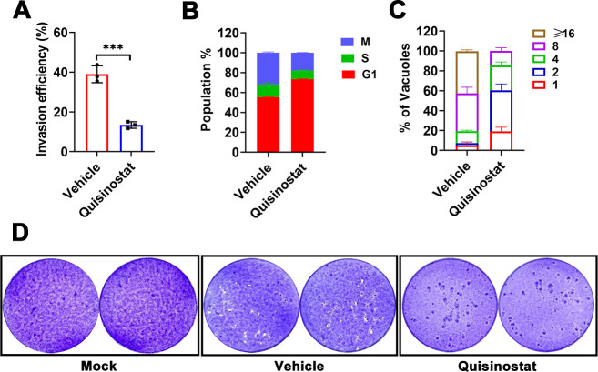
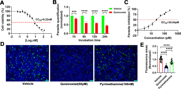
Toxoplasma gondii, an opportunistic pathogen, poses severe threats to immunocompromised individuals and fetuses of newly infected pregnant women. The current gold-standard treatment, a combination of pyrimethamine and sulfadiazine, is limited by severe adverse events, necessitating the development of novel therapeutic agents. In vitro, a CCK8 assay demonstrated that quisinostat inhibited HeLa cell proliferation in a dose-dependent manner, with a CC50 of 8.22 nM. Regarding T. gondii tachyzoites, quisinostat exhibited time-dependent inhibition of extracellular parasite activity and suppressed intracellular parasite proliferation, with an EC50 of 25.84 pM, and a high selectivity index (SI = 318.11). Quisinostat disrupted the T. gondii lytic cycle by decreasing invasion rates, inducing G1 cell cycle arrest, reducing replication, and shrinking plaque size. Ultrastructural analysis indicated that quisinostat treatment led to membrane damage, enhanced lactate dehydrogenase (LDH) release, and apoptotic cell death in tachyzoites, whereas no significant change in reactive oxygen species (ROS) levels was detected. Proteome analysis identified 77 upregulated and 205 downregulated proteins, which were enriched in functions associated with protein dephosphorylation and ion transport, as well as pathways, such as non-homologous end-joining. Molecular docking studies revealed a strong interaction between quisinostat and T. gondii HDAC3. In vivo, treatment with quisinostat increased the survival time of mice infected with virulent RH strain. In mice infected with low-virulent ME49 tachyzoites, quisinostat treatment decreased parasite burden in multiple organs and increased the survival to 80%. Taken together, these findings demonstrate that quisinostat has potent anti-Toxoplasma activity both in vitro and in vivo, which offers promise for treatment of human toxoplasmosis.
期刊介绍:
Antimicrobial Agents and Chemotherapy (AAC) features interdisciplinary studies that build our understanding of the underlying mechanisms and therapeutic applications of antimicrobial and antiparasitic agents and chemotherapy.
 求助内容:
求助内容: 应助结果提醒方式:


