Ai-Lan Nguyen, Dana Horakova, Eva H Havrdova, Michael Barnett, Maria Pia Sormani, Nicola De Stefano, Marco Battaglini, Manuela Vaneckova, Elaine Lui, Frank Gaillard, Patricia M Desmond, Hayden Prime, Mineesh Datta, Anneke van der Walt, Vilija G Jokubaitis, Femke Podevyn, Robert Zivadinov, Bianca Weinstock-Guttman, Marie B D'hooghe, Guy Nagels, Vincent Van Pesch, Guy Laureys, Liesbeth Van Hijfte, Jeannette Lechner-Scott, Francesco Patti, Edgardo Cristiano, Juan I Rojas, Diana M Sima, Wim Van Hecke, Tomas Kalincik, Helmut Butzkueven
{"title":"多位点多发性硬化症登记中脑萎缩与残疾的关系。","authors":"Ai-Lan Nguyen, Dana Horakova, Eva H Havrdova, Michael Barnett, Maria Pia Sormani, Nicola De Stefano, Marco Battaglini, Manuela Vaneckova, Elaine Lui, Frank Gaillard, Patricia M Desmond, Hayden Prime, Mineesh Datta, Anneke van der Walt, Vilija G Jokubaitis, Femke Podevyn, Robert Zivadinov, Bianca Weinstock-Guttman, Marie B D'hooghe, Guy Nagels, Vincent Van Pesch, Guy Laureys, Liesbeth Van Hijfte, Jeannette Lechner-Scott, Francesco Patti, Edgardo Cristiano, Juan I Rojas, Diana M Sima, Wim Van Hecke, Tomas Kalincik, Helmut Butzkueven","doi":"10.1136/bmjno-2025-001126","DOIUrl":null,"url":null,"abstract":"<p><strong>Background: </strong>In a retrospective multicentre cohort study, we explored the association between brain atrophy and multiple sclerosis (MS) disability using different MRI scanners and protocols at multiple sites.</p><p><strong>Methods: </strong>Relapse-onset MS patients were included if they had two clinical MRIs 12 months apart and ≥2 Expanded Disability Status Scale (EDSS) scores. Percentage brain volume change (PBVC), percentage grey matter change (PGMC), fluid-attenuated inversion recovery (FLAIR) lesion volume change, whole brain volume (BV), grey matter volume (GMV), FLAIR lesion volume and T1 hypointense lesion volume were assessed by icobrain. Disability was measured by EDSS scores and 6-month confirmed disability progression (CDP).</p><p><strong>Results: </strong>Of the 260 relapse-onset MS patients included, 204 (78%) MRI pairs were performed in the same scanner and 56 (22%) pairs were from different scanners. 93% of patients were on treatment and mean PBVC was -0.26% (±0.52). During the median follow-up of 2.8 years from the second MRI, median EDSS change was 0.0 and 12% patients experienced 6-month CDP. Cross-sectional BV and GMV at the later MRI showed a trend for association with CDP (HR 0.99; 95% CI 0.98 to 1.00; p=0.06). Only BV at the later MRI was associated with EDSS score (β -0.03, SE 0.01, p<0.001) and the rate of EDSS change over time (β -0.001, SE 0.0003, p=0.02). There was no association between longitudinal PBVC or PGMC and CDP or EDSS (p>0.05).</p><p><strong>Conclusion: </strong>In this highly treated MS cohort with low disability accrual, only cross-sectional BV showed an association with future EDSS scores, while no MRI metric predicted 6-month CDP. These findings highlight the limitations of current clinical MRI measures in predicting disability worsening in real-world settings.</p>","PeriodicalId":52754,"journal":{"name":"BMJ Neurology Open","volume":"7 2","pages":"e001126"},"PeriodicalIF":2.4000,"publicationDate":"2025-07-22","publicationTypes":"Journal Article","fieldsOfStudy":null,"isOpenAccess":false,"openAccessPdf":"https://www.ncbi.nlm.nih.gov/pmc/articles/PMC12306358/pdf/","citationCount":"0","resultStr":"{\"title\":\"Relationship between brain atrophy and disability in a multi-site multiple sclerosis registry.\",\"authors\":\"Ai-Lan Nguyen, Dana Horakova, Eva H Havrdova, Michael Barnett, Maria Pia Sormani, Nicola De Stefano, Marco Battaglini, Manuela Vaneckova, Elaine Lui, Frank Gaillard, Patricia M Desmond, Hayden Prime, Mineesh Datta, Anneke van der Walt, Vilija G Jokubaitis, Femke Podevyn, Robert Zivadinov, Bianca Weinstock-Guttman, Marie B D'hooghe, Guy Nagels, Vincent Van Pesch, Guy Laureys, Liesbeth Van Hijfte, Jeannette Lechner-Scott, Francesco Patti, Edgardo Cristiano, Juan I Rojas, Diana M Sima, Wim Van Hecke, Tomas Kalincik, Helmut Butzkueven\",\"doi\":\"10.1136/bmjno-2025-001126\",\"DOIUrl\":null,\"url\":null,\"abstract\":\"<p><strong>Background: </strong>In a retrospective multicentre cohort study, we explored the association between brain atrophy and multiple sclerosis (MS) disability using different MRI scanners and protocols at multiple sites.</p><p><strong>Methods: </strong>Relapse-onset MS patients were included if they had two clinical MRIs 12 months apart and ≥2 Expanded Disability Status Scale (EDSS) scores. Percentage brain volume change (PBVC), percentage grey matter change (PGMC), fluid-attenuated inversion recovery (FLAIR) lesion volume change, whole brain volume (BV), grey matter volume (GMV), FLAIR lesion volume and T1 hypointense lesion volume were assessed by icobrain. Disability was measured by EDSS scores and 6-month confirmed disability progression (CDP).</p><p><strong>Results: </strong>Of the 260 relapse-onset MS patients included, 204 (78%) MRI pairs were performed in the same scanner and 56 (22%) pairs were from different scanners. 93% of patients were on treatment and mean PBVC was -0.26% (±0.52). During the median follow-up of 2.8 years from the second MRI, median EDSS change was 0.0 and 12% patients experienced 6-month CDP. Cross-sectional BV and GMV at the later MRI showed a trend for association with CDP (HR 0.99; 95% CI 0.98 to 1.00; p=0.06). Only BV at the later MRI was associated with EDSS score (β -0.03, SE 0.01, p<0.001) and the rate of EDSS change over time (β -0.001, SE 0.0003, p=0.02). There was no association between longitudinal PBVC or PGMC and CDP or EDSS (p>0.05).</p><p><strong>Conclusion: </strong>In this highly treated MS cohort with low disability accrual, only cross-sectional BV showed an association with future EDSS scores, while no MRI metric predicted 6-month CDP. These findings highlight the limitations of current clinical MRI measures in predicting disability worsening in real-world settings.</p>\",\"PeriodicalId\":52754,\"journal\":{\"name\":\"BMJ Neurology Open\",\"volume\":\"7 2\",\"pages\":\"e001126\"},\"PeriodicalIF\":2.4000,\"publicationDate\":\"2025-07-22\",\"publicationTypes\":\"Journal Article\",\"fieldsOfStudy\":null,\"isOpenAccess\":false,\"openAccessPdf\":\"https://www.ncbi.nlm.nih.gov/pmc/articles/PMC12306358/pdf/\",\"citationCount\":\"0\",\"resultStr\":null,\"platform\":\"Semanticscholar\",\"paperid\":null,\"PeriodicalName\":\"BMJ Neurology Open\",\"FirstCategoryId\":\"1085\",\"ListUrlMain\":\"https://doi.org/10.1136/bmjno-2025-001126\",\"RegionNum\":0,\"RegionCategory\":null,\"ArticlePicture\":[],\"TitleCN\":null,\"AbstractTextCN\":null,\"PMCID\":null,\"EPubDate\":\"2025/1/1 0:00:00\",\"PubModel\":\"eCollection\",\"JCR\":\"Q3\",\"JCRName\":\"CLINICAL NEUROLOGY\",\"Score\":null,\"Total\":0}","platform":"Semanticscholar","paperid":null,"PeriodicalName":"BMJ Neurology Open","FirstCategoryId":"1085","ListUrlMain":"https://doi.org/10.1136/bmjno-2025-001126","RegionNum":0,"RegionCategory":null,"ArticlePicture":[],"TitleCN":null,"AbstractTextCN":null,"PMCID":null,"EPubDate":"2025/1/1 0:00:00","PubModel":"eCollection","JCR":"Q3","JCRName":"CLINICAL NEUROLOGY","Score":null,"Total":0}
引用次数: 0
摘要
背景:在一项回顾性多中心队列研究中,我们在多个地点使用不同的MRI扫描仪和方案探讨了脑萎缩与多发性硬化症(MS)残疾之间的关系。方法:两次临床mri检查间隔12个月且扩展残疾状态量表(EDSS)评分≥2分的复发性MS患者被纳入。脑容量变化百分比(PBVC)、灰质变化百分比(PGMC)、液体衰减反转恢复(FLAIR)病变体积变化、全脑体积(BV)、灰质体积(GMV)、FLAIR病变体积和T1低信号病变体积。残疾通过EDSS评分和6个月确认的残疾进展(CDP)来衡量。结果:纳入的260例复发性MS患者中,204对(78%)MRI对使用同一台扫描仪,56对(22%)MRI对使用不同的扫描仪。93%的患者接受治疗,平均PBVC为-0.26%(±0.52)。在第二次MRI的中位随访2.8年期间,EDSS的中位变化为0.0,12%的患者经历了6个月的CDP。后期MRI的横截面BV和GMV显示与CDP相关的趋势(HR 0.99;95% CI 0.98 ~ 1.00;p = 0.06)。只有后期MRI BV与EDSS评分相关(β -0.03, SE 0.01, p0.05)。结论:在这个高度治疗的低残疾累积的MS队列中,只有横断面BV显示与未来EDSS评分相关,而没有MRI指标预测6个月的CDP。这些发现强调了当前临床MRI测量在预测现实环境中残疾恶化方面的局限性。
Relationship between brain atrophy and disability in a multi-site multiple sclerosis registry.
Background: In a retrospective multicentre cohort study, we explored the association between brain atrophy and multiple sclerosis (MS) disability using different MRI scanners and protocols at multiple sites.
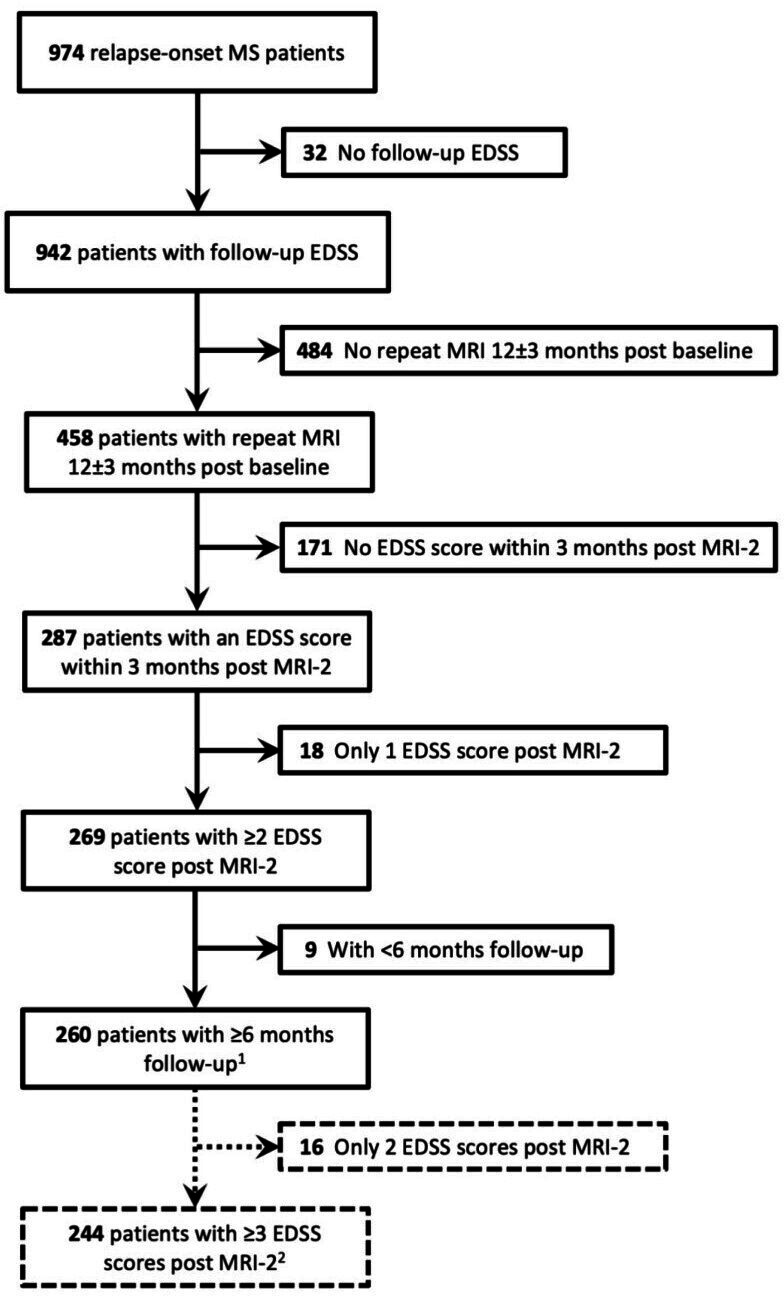
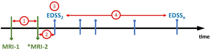
Methods: Relapse-onset MS patients were included if they had two clinical MRIs 12 months apart and ≥2 Expanded Disability Status Scale (EDSS) scores. Percentage brain volume change (PBVC), percentage grey matter change (PGMC), fluid-attenuated inversion recovery (FLAIR) lesion volume change, whole brain volume (BV), grey matter volume (GMV), FLAIR lesion volume and T1 hypointense lesion volume were assessed by icobrain. Disability was measured by EDSS scores and 6-month confirmed disability progression (CDP).
Results: Of the 260 relapse-onset MS patients included, 204 (78%) MRI pairs were performed in the same scanner and 56 (22%) pairs were from different scanners. 93% of patients were on treatment and mean PBVC was -0.26% (±0.52). During the median follow-up of 2.8 years from the second MRI, median EDSS change was 0.0 and 12% patients experienced 6-month CDP. Cross-sectional BV and GMV at the later MRI showed a trend for association with CDP (HR 0.99; 95% CI 0.98 to 1.00; p=0.06). Only BV at the later MRI was associated with EDSS score (β -0.03, SE 0.01, p<0.001) and the rate of EDSS change over time (β -0.001, SE 0.0003, p=0.02). There was no association between longitudinal PBVC or PGMC and CDP or EDSS (p>0.05).
Conclusion: In this highly treated MS cohort with low disability accrual, only cross-sectional BV showed an association with future EDSS scores, while no MRI metric predicted 6-month CDP. These findings highlight the limitations of current clinical MRI measures in predicting disability worsening in real-world settings.

 求助内容:
求助内容: 应助结果提醒方式:


