Somayeh Meysami, Saurabh Garg, Sam Hashemi, Nasrin Akbari, Ahmed Gouda, Yosef Gavriel Chodakiewitz, Thanh Duc Nguyen, Rajpaul Attariwala, Kellyann Niotis, David A Merrill, Cyrus A Raji
{"title":"吸烟可以预测10134名健康人的脑萎缩,并可能受到体重指数的影响。","authors":"Somayeh Meysami, Saurabh Garg, Sam Hashemi, Nasrin Akbari, Ahmed Gouda, Yosef Gavriel Chodakiewitz, Thanh Duc Nguyen, Rajpaul Attariwala, Kellyann Niotis, David A Merrill, Cyrus A Raji","doi":"10.1038/s44400-025-00024-0","DOIUrl":null,"url":null,"abstract":"<p><p>Cigarette smoking is a risk factor for Alzheimer's and vascular dementia, but its impact on brain volume loss, a neurodegeneration biomarker on MRI, is unclear. In total, 10,134 participants from 4 sites were scanned with a whole-body 1.5 T MRI protocol with separate dedicated structural neuroimaging with 3D T1 MPRAGE sequences. Smokers versus non-smokers were compared by gray and white matter volumes normalized to total intracranial volume using a two-tailed <i>t</i>-test. Smokers had lower normalized gray (<i>t</i> = -7.806e+00, <i>p</i> = 6.508e-15) and white matter volumes (<i>t</i> = -7.374e + 00, <i>p</i> = 1.791e-13) compared to non-smokers. Adjusting for age, sex, study site, BMI, and multiple comparisons, higher pack years of smoking predicted volume loss in such regions as total gray matter volume, total white matter volume, temporal lobe, parietal lobe, hippocampus, precuneus, and posterior cingulate. The inclusion and exclusion of BMI from the model suggested an influence of this variable.</p>","PeriodicalId":520469,"journal":{"name":"NPJ dementia","volume":"1 1","pages":"17"},"PeriodicalIF":0.0000,"publicationDate":"2025-01-01","publicationTypes":"Journal Article","fieldsOfStudy":null,"isOpenAccess":false,"openAccessPdf":"https://www.ncbi.nlm.nih.gov/pmc/articles/PMC12286857/pdf/","citationCount":"0","resultStr":"{\"title\":\"Smoking predicts brain atrophy in 10,134 healthy individuals and is potentially influenced by body mass index.\",\"authors\":\"Somayeh Meysami, Saurabh Garg, Sam Hashemi, Nasrin Akbari, Ahmed Gouda, Yosef Gavriel Chodakiewitz, Thanh Duc Nguyen, Rajpaul Attariwala, Kellyann Niotis, David A Merrill, Cyrus A Raji\",\"doi\":\"10.1038/s44400-025-00024-0\",\"DOIUrl\":null,\"url\":null,\"abstract\":\"<p><p>Cigarette smoking is a risk factor for Alzheimer's and vascular dementia, but its impact on brain volume loss, a neurodegeneration biomarker on MRI, is unclear. In total, 10,134 participants from 4 sites were scanned with a whole-body 1.5 T MRI protocol with separate dedicated structural neuroimaging with 3D T1 MPRAGE sequences. Smokers versus non-smokers were compared by gray and white matter volumes normalized to total intracranial volume using a two-tailed <i>t</i>-test. Smokers had lower normalized gray (<i>t</i> = -7.806e+00, <i>p</i> = 6.508e-15) and white matter volumes (<i>t</i> = -7.374e + 00, <i>p</i> = 1.791e-13) compared to non-smokers. Adjusting for age, sex, study site, BMI, and multiple comparisons, higher pack years of smoking predicted volume loss in such regions as total gray matter volume, total white matter volume, temporal lobe, parietal lobe, hippocampus, precuneus, and posterior cingulate. The inclusion and exclusion of BMI from the model suggested an influence of this variable.</p>\",\"PeriodicalId\":520469,\"journal\":{\"name\":\"NPJ dementia\",\"volume\":\"1 1\",\"pages\":\"17\"},\"PeriodicalIF\":0.0000,\"publicationDate\":\"2025-01-01\",\"publicationTypes\":\"Journal Article\",\"fieldsOfStudy\":null,\"isOpenAccess\":false,\"openAccessPdf\":\"https://www.ncbi.nlm.nih.gov/pmc/articles/PMC12286857/pdf/\",\"citationCount\":\"0\",\"resultStr\":null,\"platform\":\"Semanticscholar\",\"paperid\":null,\"PeriodicalName\":\"NPJ dementia\",\"FirstCategoryId\":\"1085\",\"ListUrlMain\":\"https://doi.org/10.1038/s44400-025-00024-0\",\"RegionNum\":0,\"RegionCategory\":null,\"ArticlePicture\":[],\"TitleCN\":null,\"AbstractTextCN\":null,\"PMCID\":null,\"EPubDate\":\"2025/7/23 0:00:00\",\"PubModel\":\"Epub\",\"JCR\":\"\",\"JCRName\":\"\",\"Score\":null,\"Total\":0}","platform":"Semanticscholar","paperid":null,"PeriodicalName":"NPJ dementia","FirstCategoryId":"1085","ListUrlMain":"https://doi.org/10.1038/s44400-025-00024-0","RegionNum":0,"RegionCategory":null,"ArticlePicture":[],"TitleCN":null,"AbstractTextCN":null,"PMCID":null,"EPubDate":"2025/7/23 0:00:00","PubModel":"Epub","JCR":"","JCRName":"","Score":null,"Total":0}
引用次数: 0
摘要
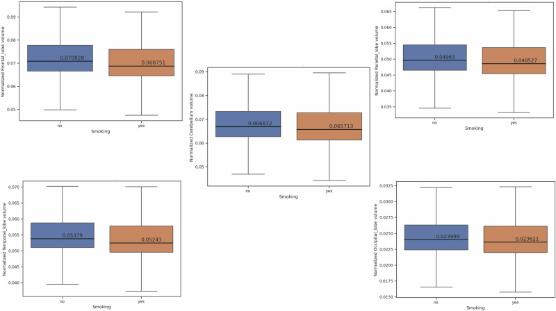
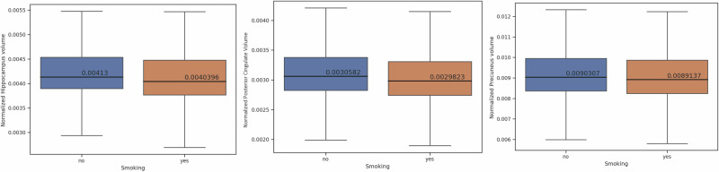
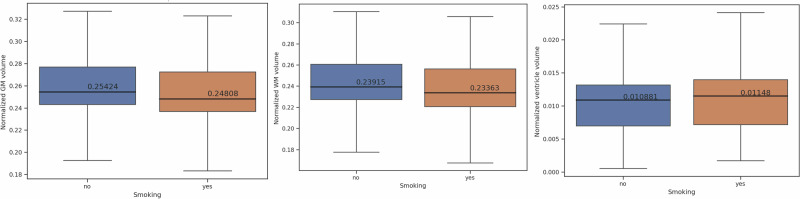
吸烟是阿尔茨海默氏症和血管性痴呆的一个危险因素,但它对脑容量损失的影响尚不清楚。脑容量损失是核磁共振成像(MRI)显示的一种神经退行性生物标志物。共对来自4个地点的10,134名参与者进行了全身1.5 T MRI扫描,并使用3D T1 MPRAGE序列进行了单独的专用结构神经成像。采用双尾t检验比较吸烟者和非吸烟者的灰质和白质体积与总颅内体积的归一化。与不吸烟者相比,吸烟者的归一化灰质(t = -7.806e+00, p = 6.508e-15)和白质体积(t = -7.374e +00, p = 1.791e-13)较低。调整年龄、性别、研究地点、BMI和多重比较后,吸烟年数越高,预测灰质总体积、白质总体积、颞叶、顶叶、海马体、钮扣前叶和后扣带等区域的体积损失。从模型中纳入和排除BMI表明了该变量的影响。
Smoking predicts brain atrophy in 10,134 healthy individuals and is potentially influenced by body mass index.
Cigarette smoking is a risk factor for Alzheimer's and vascular dementia, but its impact on brain volume loss, a neurodegeneration biomarker on MRI, is unclear. In total, 10,134 participants from 4 sites were scanned with a whole-body 1.5 T MRI protocol with separate dedicated structural neuroimaging with 3D T1 MPRAGE sequences. Smokers versus non-smokers were compared by gray and white matter volumes normalized to total intracranial volume using a two-tailed t-test. Smokers had lower normalized gray (t = -7.806e+00, p = 6.508e-15) and white matter volumes (t = -7.374e + 00, p = 1.791e-13) compared to non-smokers. Adjusting for age, sex, study site, BMI, and multiple comparisons, higher pack years of smoking predicted volume loss in such regions as total gray matter volume, total white matter volume, temporal lobe, parietal lobe, hippocampus, precuneus, and posterior cingulate. The inclusion and exclusion of BMI from the model suggested an influence of this variable.

 求助内容:
求助内容: 应助结果提醒方式:


