{"title":"脑干体积减少是阿尔茨海默病高危人群的关键宏观结构指标。","authors":"Thomas M Lancaster, Kevin Murphy, Hannah Chandler","doi":"10.1186/s13195-025-01829-0","DOIUrl":null,"url":null,"abstract":"<p><strong>Background: </strong>Alterations to brain macrostructure, assessed via T1-weighted magnetic resonance imaging are observed in preclinical models of Alzheimer's disease (AD), reflecting susceptibility, prodromal stages of AD or correlates of early AD pathophysiology. While changes in cingulate and medial temporal lobe structures may be functionally implicated in cognitive decline, little is known about the viability of brain-based biomarkers that support autonomic functions implicated in preclinical AD risk such as the brainstem.</p><p><strong>Methods: </strong>In a series of multiple linear regressions, we assess the volume of the brainstem in two asymptomatic at-AD-risk samples, assessed via the presence of either mild cognitive impairment (MCI, N = 148), or extremely high polygenic risk (N = 13) with matched demographics (mean age = 67 [range 58-76], in both cases). We further determine the strength of the association, compared to 150 other structural MRI features.</p><p><strong>Results: </strong>We observed brainstem volume reductions (MCI: b = -0.29, P = 0.018; Genetic risk: b = -1.29, P = 0.002) in both samples. The magnitude of each preclinical AD marker (MCI / AD-polygenic risk)- brainstem association was empirically larger (Z > 2.3, P < 0.05, in both cases) than 150 frequently segmented MRI features. We further replicate the negative AD-polygenic risk score- brainstem association in UK Biobank (N = 31968; b = -0.002, P = 0.03), with weaker evidence that the association was larger than all other MRI features (Z = 1.622; P = 0.052).</p><p><strong>Conclusions: </strong>These observations suggest that AD risk, assessed via the presence of MCI or extremely high AD-polygenic risk score is linked to reduced brainstem volume before most typically observed morphological brain alterations. This conforms with evidence implicating the brainstem as one of the earliest sites of morphological neurodegeneration and provides a plausible biological mechanism linking prodromal autonomic symptoms to AD risk in later life. These observations warrant future investigation into the molecular correlates of AD-linked brainstem dysfunction, assessment as a candidate biomarker, and the exploration of brainstem mediated treatment strategies in AD prevention.</p>","PeriodicalId":7516,"journal":{"name":"Alzheimer's Research & Therapy","volume":"17 1","pages":"177"},"PeriodicalIF":7.6000,"publicationDate":"2025-07-26","publicationTypes":"Journal Article","fieldsOfStudy":null,"isOpenAccess":false,"openAccessPdf":"https://www.ncbi.nlm.nih.gov/pmc/articles/PMC12296611/pdf/","citationCount":"0","resultStr":"{\"title\":\"Reductions in brainstem volume as a key macrostructural indicator in at-risk populations for Alzheimer's disease.\",\"authors\":\"Thomas M Lancaster, Kevin Murphy, Hannah Chandler\",\"doi\":\"10.1186/s13195-025-01829-0\",\"DOIUrl\":null,\"url\":null,\"abstract\":\"<p><strong>Background: </strong>Alterations to brain macrostructure, assessed via T1-weighted magnetic resonance imaging are observed in preclinical models of Alzheimer's disease (AD), reflecting susceptibility, prodromal stages of AD or correlates of early AD pathophysiology. While changes in cingulate and medial temporal lobe structures may be functionally implicated in cognitive decline, little is known about the viability of brain-based biomarkers that support autonomic functions implicated in preclinical AD risk such as the brainstem.</p><p><strong>Methods: </strong>In a series of multiple linear regressions, we assess the volume of the brainstem in two asymptomatic at-AD-risk samples, assessed via the presence of either mild cognitive impairment (MCI, N = 148), or extremely high polygenic risk (N = 13) with matched demographics (mean age = 67 [range 58-76], in both cases). We further determine the strength of the association, compared to 150 other structural MRI features.</p><p><strong>Results: </strong>We observed brainstem volume reductions (MCI: b = -0.29, P = 0.018; Genetic risk: b = -1.29, P = 0.002) in both samples. The magnitude of each preclinical AD marker (MCI / AD-polygenic risk)- brainstem association was empirically larger (Z > 2.3, P < 0.05, in both cases) than 150 frequently segmented MRI features. We further replicate the negative AD-polygenic risk score- brainstem association in UK Biobank (N = 31968; b = -0.002, P = 0.03), with weaker evidence that the association was larger than all other MRI features (Z = 1.622; P = 0.052).</p><p><strong>Conclusions: </strong>These observations suggest that AD risk, assessed via the presence of MCI or extremely high AD-polygenic risk score is linked to reduced brainstem volume before most typically observed morphological brain alterations. This conforms with evidence implicating the brainstem as one of the earliest sites of morphological neurodegeneration and provides a plausible biological mechanism linking prodromal autonomic symptoms to AD risk in later life. These observations warrant future investigation into the molecular correlates of AD-linked brainstem dysfunction, assessment as a candidate biomarker, and the exploration of brainstem mediated treatment strategies in AD prevention.</p>\",\"PeriodicalId\":7516,\"journal\":{\"name\":\"Alzheimer's Research & Therapy\",\"volume\":\"17 1\",\"pages\":\"177\"},\"PeriodicalIF\":7.6000,\"publicationDate\":\"2025-07-26\",\"publicationTypes\":\"Journal Article\",\"fieldsOfStudy\":null,\"isOpenAccess\":false,\"openAccessPdf\":\"https://www.ncbi.nlm.nih.gov/pmc/articles/PMC12296611/pdf/\",\"citationCount\":\"0\",\"resultStr\":null,\"platform\":\"Semanticscholar\",\"paperid\":null,\"PeriodicalName\":\"Alzheimer's Research & Therapy\",\"FirstCategoryId\":\"3\",\"ListUrlMain\":\"https://doi.org/10.1186/s13195-025-01829-0\",\"RegionNum\":1,\"RegionCategory\":\"医学\",\"ArticlePicture\":[],\"TitleCN\":null,\"AbstractTextCN\":null,\"PMCID\":null,\"EPubDate\":\"\",\"PubModel\":\"\",\"JCR\":\"Q1\",\"JCRName\":\"CLINICAL NEUROLOGY\",\"Score\":null,\"Total\":0}","platform":"Semanticscholar","paperid":null,"PeriodicalName":"Alzheimer's Research & Therapy","FirstCategoryId":"3","ListUrlMain":"https://doi.org/10.1186/s13195-025-01829-0","RegionNum":1,"RegionCategory":"医学","ArticlePicture":[],"TitleCN":null,"AbstractTextCN":null,"PMCID":null,"EPubDate":"","PubModel":"","JCR":"Q1","JCRName":"CLINICAL NEUROLOGY","Score":null,"Total":0}
引用次数: 0
摘要
背景:通过t1加权磁共振成像在阿尔茨海默病(AD)的临床前模型中观察到脑宏观结构的改变,反映了AD的易感性、前驱阶段或早期AD病理生理的相关因素。虽然扣带和内侧颞叶结构的变化可能在功能上与认知能力下降有关,但对于支持与临床前AD风险相关的自主神经功能(如脑干)的基于大脑的生物标志物的可行性知之甚少。方法:在一系列多元线性回归中,我们评估了两个无症状ad风险样本的脑干体积,通过轻度认知障碍(MCI, N = 148)或极高多基因风险(N = 13)的存在进行评估,并匹配人口统计学(两种情况下平均年龄= 67[范围58-76])。我们进一步确定了这种关联的强度,并与其他150个MRI结构特征进行了比较。结果:观察到脑干体积减小(MCI: b = -0.29, P = 0.018;遗传风险:b = -1.29, P = 0.002)。每个临床前AD标志物(MCI / AD多基因风险)与脑干的关联程度在经验上更大(z> 2.3, P)。结论:这些观察结果表明,通过MCI或极高AD多基因风险评分评估的AD风险与大多数典型观察到的脑形态学改变之前脑干体积减少有关。这与脑干作为形态学神经退行性变的最早部位之一的证据一致,并提供了将前驱自主神经症状与晚年阿尔茨海默病风险联系起来的似是而非的生物学机制。这些观察结果为进一步研究AD相关脑干功能障碍的分子相关性、作为候选生物标志物的评估以及探索脑干介导的AD预防治疗策略提供了依据。
Reductions in brainstem volume as a key macrostructural indicator in at-risk populations for Alzheimer's disease.
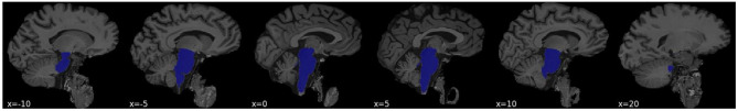
Background: Alterations to brain macrostructure, assessed via T1-weighted magnetic resonance imaging are observed in preclinical models of Alzheimer's disease (AD), reflecting susceptibility, prodromal stages of AD or correlates of early AD pathophysiology. While changes in cingulate and medial temporal lobe structures may be functionally implicated in cognitive decline, little is known about the viability of brain-based biomarkers that support autonomic functions implicated in preclinical AD risk such as the brainstem.
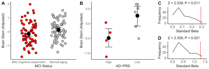
Methods: In a series of multiple linear regressions, we assess the volume of the brainstem in two asymptomatic at-AD-risk samples, assessed via the presence of either mild cognitive impairment (MCI, N = 148), or extremely high polygenic risk (N = 13) with matched demographics (mean age = 67 [range 58-76], in both cases). We further determine the strength of the association, compared to 150 other structural MRI features.
Results: We observed brainstem volume reductions (MCI: b = -0.29, P = 0.018; Genetic risk: b = -1.29, P = 0.002) in both samples. The magnitude of each preclinical AD marker (MCI / AD-polygenic risk)- brainstem association was empirically larger (Z > 2.3, P < 0.05, in both cases) than 150 frequently segmented MRI features. We further replicate the negative AD-polygenic risk score- brainstem association in UK Biobank (N = 31968; b = -0.002, P = 0.03), with weaker evidence that the association was larger than all other MRI features (Z = 1.622; P = 0.052).
Conclusions: These observations suggest that AD risk, assessed via the presence of MCI or extremely high AD-polygenic risk score is linked to reduced brainstem volume before most typically observed morphological brain alterations. This conforms with evidence implicating the brainstem as one of the earliest sites of morphological neurodegeneration and provides a plausible biological mechanism linking prodromal autonomic symptoms to AD risk in later life. These observations warrant future investigation into the molecular correlates of AD-linked brainstem dysfunction, assessment as a candidate biomarker, and the exploration of brainstem mediated treatment strategies in AD prevention.
期刊介绍:
Alzheimer's Research & Therapy is an international peer-reviewed journal that focuses on translational research into Alzheimer's disease and other neurodegenerative diseases. It publishes open-access basic research, clinical trials, drug discovery and development studies, and epidemiologic studies. The journal also includes reviews, viewpoints, commentaries, debates, and reports. All articles published in Alzheimer's Research & Therapy are included in several reputable databases such as CAS, Current contents, DOAJ, Embase, Journal Citation Reports/Science Edition, MEDLINE, PubMed, PubMed Central, Science Citation Index Expanded (Web of Science) and Scopus.

 求助内容:
求助内容: 应助结果提醒方式:


