Anna Szelenyi, Philipp Stelzer, Christian Wassipaul, Jakob Kittinger, Andreas Strassl, Victor Schmidbauer, Martin Luther Watzenböck, Florian Lindenlaub, Michael Arnoldner, Michael Weber, Matthias Pinter, Ruxandra-Iulia Milos, Dietmar Tamandl
{"title":"基于光子计数检测器的肝细胞癌ct成像参数优化——重建核和层厚的影响。","authors":"Anna Szelenyi, Philipp Stelzer, Christian Wassipaul, Jakob Kittinger, Andreas Strassl, Victor Schmidbauer, Martin Luther Watzenböck, Florian Lindenlaub, Michael Arnoldner, Michael Weber, Matthias Pinter, Ruxandra-Iulia Milos, Dietmar Tamandl","doi":"10.3390/tomography11070077","DOIUrl":null,"url":null,"abstract":"<p><strong>Background: </strong>The use of photon-counting detector computed tomography (PCD-CT) has improved image quality in cardiac, pulmonary, and musculoskeletal imaging. Abdominal imaging research, especially about the use of PCD-CT in hepatocellular carcinoma (HCC), is sparse.</p><p><strong>Objectives: </strong>We aimed to compare the image quality of tumors, the liver parenchyma, and the vasculature in patients with HCC using PCD-CT reconstructions at different slice thicknesses and kernels to identify the most appropriate settings for the clinical routine.</p><p><strong>Methods: </strong>CT exams from twenty adult patients with HCC performed with a clinically approved, first-generation PCD-CT scanner (Naeotom Alpha<sup>®</sup>, Siemens Healthineers), were retrospectively reviewed. For each patient, images were reconstructed at four different sharp kernels, designed for abdominal imaging (Br40; Br44; Br48; Br56) and at three slice thicknesses (0.4 mm; 1 mm; 3 mm). The reconstruction with the Br40 kernel at 3 mm (Br40<sub>3 mm</sub>) was used as a clinical reference. Three readers independently assessed the image quality of different anatomical abdominal structures and hypervascular HCC lesions using a five-point Likert scale. In addition, image sharpness was assessed using line-density profiles.</p><p><strong>Results: </strong>Compared with the clinical reference, the Br44<sub>1 mm</sub> and Br48<sub>1 mm</sub> reconstructions were rated superior for the assessment of the hepatic vasculature (median difference +0.67 [+0.33 to +1.33], <i>p</i> < 0.001 and +1.00 [+0.67 to +1.67], <i>p</i> < 0.001). Reconstructions for Br40<sub>1 mm</sub> (+0.33 [-0.67 to +1.00], <i>p</i> < 0.001), and Br44<sub>3 mm</sub> (+0.0 [0.0 to +1.00], <i>p</i> = 0.030) were scored superior for overall image quality. The noise demonstrated a continuous increase when using sharper kernels and thinner slices than Br40<sub>3 mm</sub> (<i>p</i> < 0.001), leading to a decrease in contrast-to-noise ratio. Although there was a trend toward increased image sharpness using the slope analysis with higher kernels, this was not significantly different compared with the reference standard.</p><p><strong>Conclusion: </strong>PCD-CT reconstruction Br40<sub>1 mm</sub> was the most suitable setting for overall image quality, while reconstructions with sharper kernels (Br44<sub>1 mm</sub> and Br48<sub>1 mm</sub>) can be considered for the assessment of the hepatic vasculature in patients with HCC.</p>","PeriodicalId":51330,"journal":{"name":"Tomography","volume":"11 7","pages":""},"PeriodicalIF":2.2000,"publicationDate":"2025-06-27","publicationTypes":"Journal Article","fieldsOfStudy":null,"isOpenAccess":false,"openAccessPdf":"https://www.ncbi.nlm.nih.gov/pmc/articles/PMC12299250/pdf/","citationCount":"0","resultStr":"{\"title\":\"Optimizing Imaging Parameters for Assessment of Hepatocellular Carcinoma Using Photon-Counting Detector Computed Tomography-Impact of Reconstruction Kernel and Slice Thickness.\",\"authors\":\"Anna Szelenyi, Philipp Stelzer, Christian Wassipaul, Jakob Kittinger, Andreas Strassl, Victor Schmidbauer, Martin Luther Watzenböck, Florian Lindenlaub, Michael Arnoldner, Michael Weber, Matthias Pinter, Ruxandra-Iulia Milos, Dietmar Tamandl\",\"doi\":\"10.3390/tomography11070077\",\"DOIUrl\":null,\"url\":null,\"abstract\":\"<p><strong>Background: </strong>The use of photon-counting detector computed tomography (PCD-CT) has improved image quality in cardiac, pulmonary, and musculoskeletal imaging. Abdominal imaging research, especially about the use of PCD-CT in hepatocellular carcinoma (HCC), is sparse.</p><p><strong>Objectives: </strong>We aimed to compare the image quality of tumors, the liver parenchyma, and the vasculature in patients with HCC using PCD-CT reconstructions at different slice thicknesses and kernels to identify the most appropriate settings for the clinical routine.</p><p><strong>Methods: </strong>CT exams from twenty adult patients with HCC performed with a clinically approved, first-generation PCD-CT scanner (Naeotom Alpha<sup>®</sup>, Siemens Healthineers), were retrospectively reviewed. For each patient, images were reconstructed at four different sharp kernels, designed for abdominal imaging (Br40; Br44; Br48; Br56) and at three slice thicknesses (0.4 mm; 1 mm; 3 mm). The reconstruction with the Br40 kernel at 3 mm (Br40<sub>3 mm</sub>) was used as a clinical reference. Three readers independently assessed the image quality of different anatomical abdominal structures and hypervascular HCC lesions using a five-point Likert scale. In addition, image sharpness was assessed using line-density profiles.</p><p><strong>Results: </strong>Compared with the clinical reference, the Br44<sub>1 mm</sub> and Br48<sub>1 mm</sub> reconstructions were rated superior for the assessment of the hepatic vasculature (median difference +0.67 [+0.33 to +1.33], <i>p</i> < 0.001 and +1.00 [+0.67 to +1.67], <i>p</i> < 0.001). Reconstructions for Br40<sub>1 mm</sub> (+0.33 [-0.67 to +1.00], <i>p</i> < 0.001), and Br44<sub>3 mm</sub> (+0.0 [0.0 to +1.00], <i>p</i> = 0.030) were scored superior for overall image quality. The noise demonstrated a continuous increase when using sharper kernels and thinner slices than Br40<sub>3 mm</sub> (<i>p</i> < 0.001), leading to a decrease in contrast-to-noise ratio. Although there was a trend toward increased image sharpness using the slope analysis with higher kernels, this was not significantly different compared with the reference standard.</p><p><strong>Conclusion: </strong>PCD-CT reconstruction Br40<sub>1 mm</sub> was the most suitable setting for overall image quality, while reconstructions with sharper kernels (Br44<sub>1 mm</sub> and Br48<sub>1 mm</sub>) can be considered for the assessment of the hepatic vasculature in patients with HCC.</p>\",\"PeriodicalId\":51330,\"journal\":{\"name\":\"Tomography\",\"volume\":\"11 7\",\"pages\":\"\"},\"PeriodicalIF\":2.2000,\"publicationDate\":\"2025-06-27\",\"publicationTypes\":\"Journal Article\",\"fieldsOfStudy\":null,\"isOpenAccess\":false,\"openAccessPdf\":\"https://www.ncbi.nlm.nih.gov/pmc/articles/PMC12299250/pdf/\",\"citationCount\":\"0\",\"resultStr\":null,\"platform\":\"Semanticscholar\",\"paperid\":null,\"PeriodicalName\":\"Tomography\",\"FirstCategoryId\":\"3\",\"ListUrlMain\":\"https://doi.org/10.3390/tomography11070077\",\"RegionNum\":4,\"RegionCategory\":\"医学\",\"ArticlePicture\":[],\"TitleCN\":null,\"AbstractTextCN\":null,\"PMCID\":null,\"EPubDate\":\"\",\"PubModel\":\"\",\"JCR\":\"Q2\",\"JCRName\":\"RADIOLOGY, NUCLEAR MEDICINE & MEDICAL IMAGING\",\"Score\":null,\"Total\":0}","platform":"Semanticscholar","paperid":null,"PeriodicalName":"Tomography","FirstCategoryId":"3","ListUrlMain":"https://doi.org/10.3390/tomography11070077","RegionNum":4,"RegionCategory":"医学","ArticlePicture":[],"TitleCN":null,"AbstractTextCN":null,"PMCID":null,"EPubDate":"","PubModel":"","JCR":"Q2","JCRName":"RADIOLOGY, NUCLEAR MEDICINE & MEDICAL IMAGING","Score":null,"Total":0}
Optimizing Imaging Parameters for Assessment of Hepatocellular Carcinoma Using Photon-Counting Detector Computed Tomography-Impact of Reconstruction Kernel and Slice Thickness.
Background: The use of photon-counting detector computed tomography (PCD-CT) has improved image quality in cardiac, pulmonary, and musculoskeletal imaging. Abdominal imaging research, especially about the use of PCD-CT in hepatocellular carcinoma (HCC), is sparse.
Objectives: We aimed to compare the image quality of tumors, the liver parenchyma, and the vasculature in patients with HCC using PCD-CT reconstructions at different slice thicknesses and kernels to identify the most appropriate settings for the clinical routine.
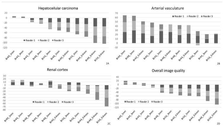
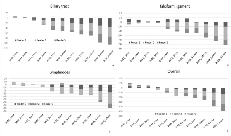
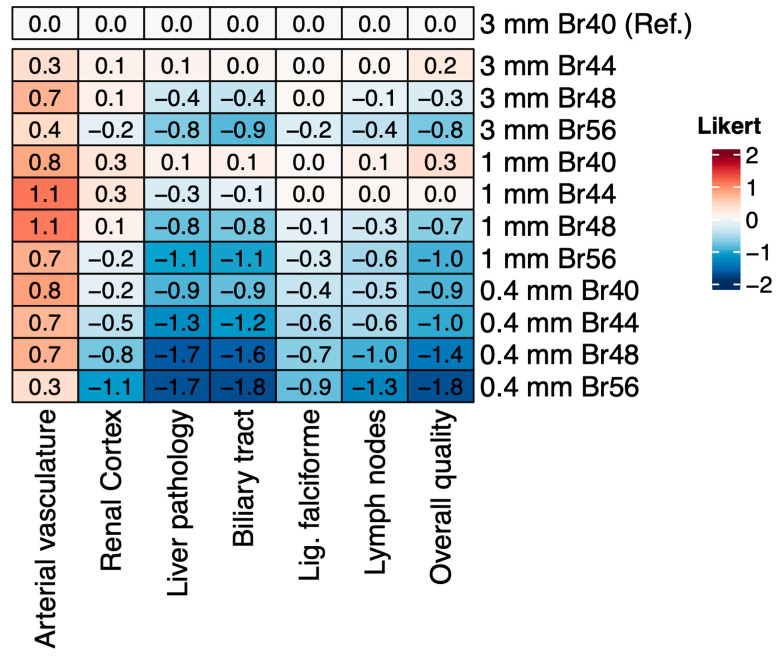
Methods: CT exams from twenty adult patients with HCC performed with a clinically approved, first-generation PCD-CT scanner (Naeotom Alpha®, Siemens Healthineers), were retrospectively reviewed. For each patient, images were reconstructed at four different sharp kernels, designed for abdominal imaging (Br40; Br44; Br48; Br56) and at three slice thicknesses (0.4 mm; 1 mm; 3 mm). The reconstruction with the Br40 kernel at 3 mm (Br403 mm) was used as a clinical reference. Three readers independently assessed the image quality of different anatomical abdominal structures and hypervascular HCC lesions using a five-point Likert scale. In addition, image sharpness was assessed using line-density profiles.
Results: Compared with the clinical reference, the Br441 mm and Br481 mm reconstructions were rated superior for the assessment of the hepatic vasculature (median difference +0.67 [+0.33 to +1.33], p < 0.001 and +1.00 [+0.67 to +1.67], p < 0.001). Reconstructions for Br401 mm (+0.33 [-0.67 to +1.00], p < 0.001), and Br443 mm (+0.0 [0.0 to +1.00], p = 0.030) were scored superior for overall image quality. The noise demonstrated a continuous increase when using sharper kernels and thinner slices than Br403 mm (p < 0.001), leading to a decrease in contrast-to-noise ratio. Although there was a trend toward increased image sharpness using the slope analysis with higher kernels, this was not significantly different compared with the reference standard.
Conclusion: PCD-CT reconstruction Br401 mm was the most suitable setting for overall image quality, while reconstructions with sharper kernels (Br441 mm and Br481 mm) can be considered for the assessment of the hepatic vasculature in patients with HCC.
TomographyMedicine-Radiology, Nuclear Medicine and Imaging
CiteScore
2.70
自引率
10.50%
发文量
222
期刊介绍:
TomographyTM publishes basic (technical and pre-clinical) and clinical scientific articles which involve the advancement of imaging technologies. Tomography encompasses studies that use single or multiple imaging modalities including for example CT, US, PET, SPECT, MR and hyperpolarization technologies, as well as optical modalities (i.e. bioluminescence, photoacoustic, endomicroscopy, fiber optic imaging and optical computed tomography) in basic sciences, engineering, preclinical and clinical medicine.
Tomography also welcomes studies involving exploration and refinement of contrast mechanisms and image-derived metrics within and across modalities toward the development of novel imaging probes for image-based feedback and intervention. The use of imaging in biology and medicine provides unparalleled opportunities to noninvasively interrogate tissues to obtain real-time dynamic and quantitative information required for diagnosis and response to interventions and to follow evolving pathological conditions. As multi-modal studies and the complexities of imaging technologies themselves are ever increasing to provide advanced information to scientists and clinicians.
Tomography provides a unique publication venue allowing investigators the opportunity to more precisely communicate integrated findings related to the diverse and heterogeneous features associated with underlying anatomical, physiological, functional, metabolic and molecular genetic activities of normal and diseased tissue. Thus Tomography publishes peer-reviewed articles which involve the broad use of imaging of any tissue and disease type including both preclinical and clinical investigations. In addition, hardware/software along with chemical and molecular probe advances are welcome as they are deemed to significantly contribute towards the long-term goal of improving the overall impact of imaging on scientific and clinical discovery.

 求助内容:
求助内容: 应助结果提醒方式:


