Emily Akerman, Daniel Aston, Eva A Rog-Zielinska, Barry Boland, Ulrich Schotten, Sander Verheule, Rebecca A Capel, Rebecca A B Burton
{"title":"心房颗粒作为酸性钙储存在心肌细胞中。","authors":"Emily Akerman, Daniel Aston, Eva A Rog-Zielinska, Barry Boland, Ulrich Schotten, Sander Verheule, Rebecca A Capel, Rebecca A B Burton","doi":"10.1093/ehjopen/oeaf083","DOIUrl":null,"url":null,"abstract":"<p><p>Acidic calcium stores significantly influence basal calcium transient amplitude and β-adrenergic responses in cardiomyocytes. Atrial myocytes contain atrial granules (AGs), small acidic organelles that store and secrete atrial natriuretic peptide (ANP) and are absent in healthy ventricular myocytes. AGs are known to be acidic and calcium-rich, but their number and location relative to other signalling sites remain unexplored. Labelling of acidic organelles in adult guinea pig cardiomyocytes showed the presence of acidic puncta throughout the cytosol. Atrial myocytes exhibited an increased concentration of acidic organelles at the nuclear poles. Live cell fluorescent studies using 4-phenyl-3-butenoic acid (PBA) to inhibit peptidylglycine α-amidating monooxygenase, a crucial component of AGs membranes, effectively eliminated staining at the nuclear poles and most acidic puncta in atrial cells, but not in ventricular cells. Our immunofluorescent labelling also emphasizes the differences in acidic punctae between atrial and ventricular myocytes by showing minimal co-localization between AG-specific ANP and lysosomal-associated membrane protein. Electron microscopy studies on goat atrial fibrillation (AF) and sham control tissue allowed visualization of AGs. Quantitative analysis revealed that AGs were positioned significantly further away from the nearest sarcoplasmic reticulum and were closer to mitochondria in AF compared to sinus rhythm control tissue. We raise the question whether the positioning of AGs is strategic for communication with other calcium-containing organelles.</p>","PeriodicalId":93995,"journal":{"name":"European heart journal open","volume":"5 4","pages":"oeaf083"},"PeriodicalIF":0.0000,"publicationDate":"2025-07-04","publicationTypes":"Journal Article","fieldsOfStudy":null,"isOpenAccess":false,"openAccessPdf":"https://www.ncbi.nlm.nih.gov/pmc/articles/PMC12284475/pdf/","citationCount":"0","resultStr":"{\"title\":\"Atrial granules as acidic calcium stores in cardiomyocytes.\",\"authors\":\"Emily Akerman, Daniel Aston, Eva A Rog-Zielinska, Barry Boland, Ulrich Schotten, Sander Verheule, Rebecca A Capel, Rebecca A B Burton\",\"doi\":\"10.1093/ehjopen/oeaf083\",\"DOIUrl\":null,\"url\":null,\"abstract\":\"<p><p>Acidic calcium stores significantly influence basal calcium transient amplitude and β-adrenergic responses in cardiomyocytes. Atrial myocytes contain atrial granules (AGs), small acidic organelles that store and secrete atrial natriuretic peptide (ANP) and are absent in healthy ventricular myocytes. AGs are known to be acidic and calcium-rich, but their number and location relative to other signalling sites remain unexplored. Labelling of acidic organelles in adult guinea pig cardiomyocytes showed the presence of acidic puncta throughout the cytosol. Atrial myocytes exhibited an increased concentration of acidic organelles at the nuclear poles. Live cell fluorescent studies using 4-phenyl-3-butenoic acid (PBA) to inhibit peptidylglycine α-amidating monooxygenase, a crucial component of AGs membranes, effectively eliminated staining at the nuclear poles and most acidic puncta in atrial cells, but not in ventricular cells. Our immunofluorescent labelling also emphasizes the differences in acidic punctae between atrial and ventricular myocytes by showing minimal co-localization between AG-specific ANP and lysosomal-associated membrane protein. Electron microscopy studies on goat atrial fibrillation (AF) and sham control tissue allowed visualization of AGs. Quantitative analysis revealed that AGs were positioned significantly further away from the nearest sarcoplasmic reticulum and were closer to mitochondria in AF compared to sinus rhythm control tissue. We raise the question whether the positioning of AGs is strategic for communication with other calcium-containing organelles.</p>\",\"PeriodicalId\":93995,\"journal\":{\"name\":\"European heart journal open\",\"volume\":\"5 4\",\"pages\":\"oeaf083\"},\"PeriodicalIF\":0.0000,\"publicationDate\":\"2025-07-04\",\"publicationTypes\":\"Journal Article\",\"fieldsOfStudy\":null,\"isOpenAccess\":false,\"openAccessPdf\":\"https://www.ncbi.nlm.nih.gov/pmc/articles/PMC12284475/pdf/\",\"citationCount\":\"0\",\"resultStr\":null,\"platform\":\"Semanticscholar\",\"paperid\":null,\"PeriodicalName\":\"European heart journal open\",\"FirstCategoryId\":\"1085\",\"ListUrlMain\":\"https://doi.org/10.1093/ehjopen/oeaf083\",\"RegionNum\":0,\"RegionCategory\":null,\"ArticlePicture\":[],\"TitleCN\":null,\"AbstractTextCN\":null,\"PMCID\":null,\"EPubDate\":\"2025/7/1 0:00:00\",\"PubModel\":\"eCollection\",\"JCR\":\"\",\"JCRName\":\"\",\"Score\":null,\"Total\":0}","platform":"Semanticscholar","paperid":null,"PeriodicalName":"European heart journal open","FirstCategoryId":"1085","ListUrlMain":"https://doi.org/10.1093/ehjopen/oeaf083","RegionNum":0,"RegionCategory":null,"ArticlePicture":[],"TitleCN":null,"AbstractTextCN":null,"PMCID":null,"EPubDate":"2025/7/1 0:00:00","PubModel":"eCollection","JCR":"","JCRName":"","Score":null,"Total":0}
Atrial granules as acidic calcium stores in cardiomyocytes.
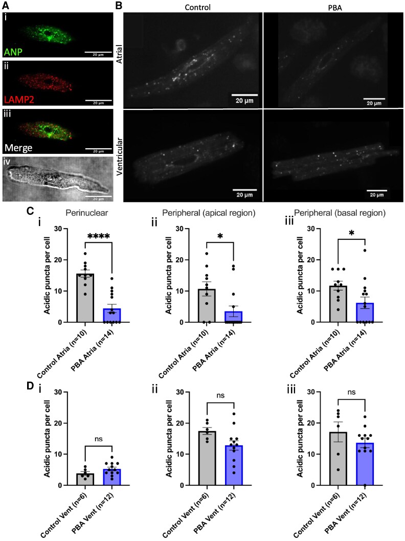
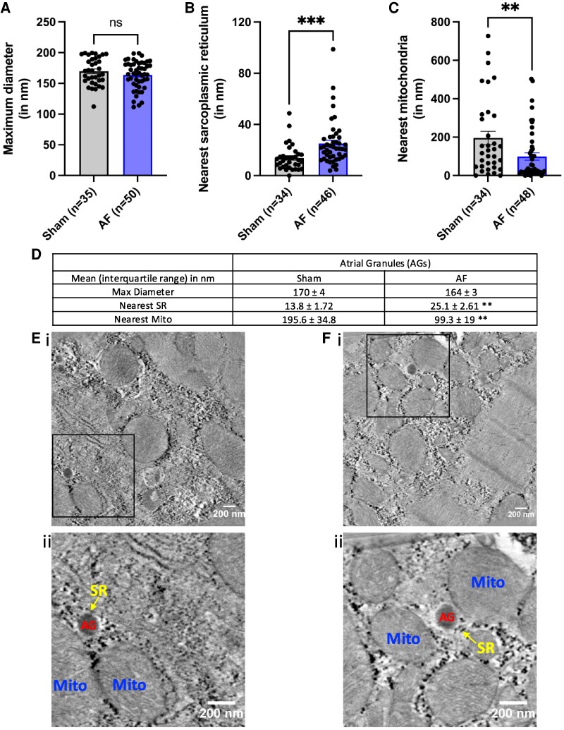
Acidic calcium stores significantly influence basal calcium transient amplitude and β-adrenergic responses in cardiomyocytes. Atrial myocytes contain atrial granules (AGs), small acidic organelles that store and secrete atrial natriuretic peptide (ANP) and are absent in healthy ventricular myocytes. AGs are known to be acidic and calcium-rich, but their number and location relative to other signalling sites remain unexplored. Labelling of acidic organelles in adult guinea pig cardiomyocytes showed the presence of acidic puncta throughout the cytosol. Atrial myocytes exhibited an increased concentration of acidic organelles at the nuclear poles. Live cell fluorescent studies using 4-phenyl-3-butenoic acid (PBA) to inhibit peptidylglycine α-amidating monooxygenase, a crucial component of AGs membranes, effectively eliminated staining at the nuclear poles and most acidic puncta in atrial cells, but not in ventricular cells. Our immunofluorescent labelling also emphasizes the differences in acidic punctae between atrial and ventricular myocytes by showing minimal co-localization between AG-specific ANP and lysosomal-associated membrane protein. Electron microscopy studies on goat atrial fibrillation (AF) and sham control tissue allowed visualization of AGs. Quantitative analysis revealed that AGs were positioned significantly further away from the nearest sarcoplasmic reticulum and were closer to mitochondria in AF compared to sinus rhythm control tissue. We raise the question whether the positioning of AGs is strategic for communication with other calcium-containing organelles.


 求助内容:
求助内容: 应助结果提醒方式:


