Shu-Xian Wu, Xiao-Long Liang, Qin-Qin Zhu, Wei Wang, Li Jiang, Huan-Huan Chen, Shui Tian, Ming Qi
{"title":"2型糖尿病患者海马亚区体积的改变与记忆和执行功能有关。","authors":"Shu-Xian Wu, Xiao-Long Liang, Qin-Qin Zhu, Wei Wang, Li Jiang, Huan-Huan Chen, Shui Tian, Ming Qi","doi":"10.4239/wjd.v16.i7.104424","DOIUrl":null,"url":null,"abstract":"<p><strong>Background: </strong>Increasing evidence has shown that hippocampal damage serves as a marker of early cognitive decline in patients with type 2 diabetes mellitus (T2DM); however, the association between hippocampal subregion volume changes and cognitive decline in different dimensions remains unclear.</p><p><strong>Aim: </strong>To investigate changes in hippocampal subregion volumes in patients with T2DM and their relationship with cognitive function impairment.</p><p><strong>Methods: </strong>Sixty patients with T2DM and 32 healthy controls were recruited. All participants underwent a 3.0 T magnetic resonance scan and a series of clinical assessments. Hippocampal subfield volumes were determined using FreeSurfer 7.4.1. A two-sample <i>t</i>-test was used to evaluate group differences. Partial correlation analysis was performed to assess the relationship between hippocampal subregion volumes and cognitive function. <sup>a</sup> <i>P</i> < 0.05 was considered statistically significant.</p><p><strong>Results: </strong>Compared with controls, the volume of right hippocampus-amygdala transition area (<i>t</i> = -3.053, <i>P</i> = 0.003) in patients with T2DM was significantly reduced, which was negatively correlated with the required time of the Trail Making Test (TMT)-A (<i>r</i> = -0.331, <i>P</i> = 0.028) and TMT-B (<i>r</i> = -0.402, <i>P</i> = 0.007) and positively correlated with the scores of Symbol Digit Modalities Test (<i>r</i> = 0.381, <i>P</i> = 0.011), Auditory Verbal Learning Test (AVLT)-N7 (<i>r</i> = 0.309, <i>P</i> = 0.041), and Digital Span Test (<i>r</i> = 0.300, <i>P</i> = 0.048). The volume of the right molecular layer (<i>t</i> = -2.998, <i>P</i> = 0.004) was also significantly reduced, which was positively associated with the scores of AVLT-N7 (<i>r</i> = 0.311, <i>P</i> = 0.045). In addition, the left hippocampal fissure volume (<i>t</i> = 3.617, <i>P</i> = 0.002) was significantly increased in patients with T2DM.</p><p><strong>Conclusion: </strong>Declines in cognitive performance, especially memory and executive function, are linked to changes in the volumes of the right hippocampus-amygdala transition area and right molecular layer in patients with T2DM.</p>","PeriodicalId":48607,"journal":{"name":"World Journal of Diabetes","volume":"16 7","pages":"104424"},"PeriodicalIF":4.6000,"publicationDate":"2025-07-15","publicationTypes":"Journal Article","fieldsOfStudy":null,"isOpenAccess":false,"openAccessPdf":"https://www.ncbi.nlm.nih.gov/pmc/articles/PMC12278078/pdf/","citationCount":"0","resultStr":"{\"title\":\"Altered hippocampal subfield volumes are associated with memory and executive function in patients with type 2 diabetes mellitus.\",\"authors\":\"Shu-Xian Wu, Xiao-Long Liang, Qin-Qin Zhu, Wei Wang, Li Jiang, Huan-Huan Chen, Shui Tian, Ming Qi\",\"doi\":\"10.4239/wjd.v16.i7.104424\",\"DOIUrl\":null,\"url\":null,\"abstract\":\"<p><strong>Background: </strong>Increasing evidence has shown that hippocampal damage serves as a marker of early cognitive decline in patients with type 2 diabetes mellitus (T2DM); however, the association between hippocampal subregion volume changes and cognitive decline in different dimensions remains unclear.</p><p><strong>Aim: </strong>To investigate changes in hippocampal subregion volumes in patients with T2DM and their relationship with cognitive function impairment.</p><p><strong>Methods: </strong>Sixty patients with T2DM and 32 healthy controls were recruited. All participants underwent a 3.0 T magnetic resonance scan and a series of clinical assessments. Hippocampal subfield volumes were determined using FreeSurfer 7.4.1. A two-sample <i>t</i>-test was used to evaluate group differences. Partial correlation analysis was performed to assess the relationship between hippocampal subregion volumes and cognitive function. <sup>a</sup> <i>P</i> < 0.05 was considered statistically significant.</p><p><strong>Results: </strong>Compared with controls, the volume of right hippocampus-amygdala transition area (<i>t</i> = -3.053, <i>P</i> = 0.003) in patients with T2DM was significantly reduced, which was negatively correlated with the required time of the Trail Making Test (TMT)-A (<i>r</i> = -0.331, <i>P</i> = 0.028) and TMT-B (<i>r</i> = -0.402, <i>P</i> = 0.007) and positively correlated with the scores of Symbol Digit Modalities Test (<i>r</i> = 0.381, <i>P</i> = 0.011), Auditory Verbal Learning Test (AVLT)-N7 (<i>r</i> = 0.309, <i>P</i> = 0.041), and Digital Span Test (<i>r</i> = 0.300, <i>P</i> = 0.048). The volume of the right molecular layer (<i>t</i> = -2.998, <i>P</i> = 0.004) was also significantly reduced, which was positively associated with the scores of AVLT-N7 (<i>r</i> = 0.311, <i>P</i> = 0.045). In addition, the left hippocampal fissure volume (<i>t</i> = 3.617, <i>P</i> = 0.002) was significantly increased in patients with T2DM.</p><p><strong>Conclusion: </strong>Declines in cognitive performance, especially memory and executive function, are linked to changes in the volumes of the right hippocampus-amygdala transition area and right molecular layer in patients with T2DM.</p>\",\"PeriodicalId\":48607,\"journal\":{\"name\":\"World Journal of Diabetes\",\"volume\":\"16 7\",\"pages\":\"104424\"},\"PeriodicalIF\":4.6000,\"publicationDate\":\"2025-07-15\",\"publicationTypes\":\"Journal Article\",\"fieldsOfStudy\":null,\"isOpenAccess\":false,\"openAccessPdf\":\"https://www.ncbi.nlm.nih.gov/pmc/articles/PMC12278078/pdf/\",\"citationCount\":\"0\",\"resultStr\":null,\"platform\":\"Semanticscholar\",\"paperid\":null,\"PeriodicalName\":\"World Journal of Diabetes\",\"FirstCategoryId\":\"3\",\"ListUrlMain\":\"https://doi.org/10.4239/wjd.v16.i7.104424\",\"RegionNum\":3,\"RegionCategory\":\"医学\",\"ArticlePicture\":[],\"TitleCN\":null,\"AbstractTextCN\":null,\"PMCID\":null,\"EPubDate\":\"\",\"PubModel\":\"\",\"JCR\":\"Q1\",\"JCRName\":\"ENDOCRINOLOGY & METABOLISM\",\"Score\":null,\"Total\":0}","platform":"Semanticscholar","paperid":null,"PeriodicalName":"World Journal of Diabetes","FirstCategoryId":"3","ListUrlMain":"https://doi.org/10.4239/wjd.v16.i7.104424","RegionNum":3,"RegionCategory":"医学","ArticlePicture":[],"TitleCN":null,"AbstractTextCN":null,"PMCID":null,"EPubDate":"","PubModel":"","JCR":"Q1","JCRName":"ENDOCRINOLOGY & METABOLISM","Score":null,"Total":0}
引用次数: 0
摘要
背景:越来越多的证据表明,海马损伤是2型糖尿病(T2DM)患者早期认知能力下降的标志;然而,海马亚区体积变化与不同维度认知能力下降之间的关系尚不清楚。目的:探讨2型糖尿病患者海马亚区体积的变化及其与认知功能障碍的关系。方法:选取T2DM患者60例,健康对照32例。所有参与者都进行了3.0 T磁共振扫描和一系列临床评估。使用FreeSurfer 7.4.1测定海马子区体积。采用双样本t检验评价组间差异。采用偏相关分析评估海马亚区体积与认知功能之间的关系。P < 0.05为差异有统计学意义。结果:与控制相比,对hippocampus-amygdala过渡区域的体积(t = -3.053, P = 0.003)患者2型糖尿病显著降低,这是负相关所需时间的轨迹测试(TMT)——(r = -0.331, P = 0.028)和TMT-B (r = -0.402, P = 0.007)和数字象征模式测试与成绩呈正相关(r = 0.381, P = 0.011),听觉言语学习测试(AVLT) -N7 (r = 0.309, P = 0.041),和数字广度测试(r = 0.300,P = 0.048)。右分子层体积(t = -2.998, P = 0.004)也显著减小,与AVLT-N7评分呈正相关(r = 0.311, P = 0.045)。此外,T2DM患者左侧海马裂体积(t = 3.617, P = 0.002)显著增加。结论:T2DM患者认知能力的下降,尤其是记忆和执行功能的下降与右侧海马-杏仁核过渡区和右侧分子层体积的变化有关。
Altered hippocampal subfield volumes are associated with memory and executive function in patients with type 2 diabetes mellitus.
Background: Increasing evidence has shown that hippocampal damage serves as a marker of early cognitive decline in patients with type 2 diabetes mellitus (T2DM); however, the association between hippocampal subregion volume changes and cognitive decline in different dimensions remains unclear.
Aim: To investigate changes in hippocampal subregion volumes in patients with T2DM and their relationship with cognitive function impairment.
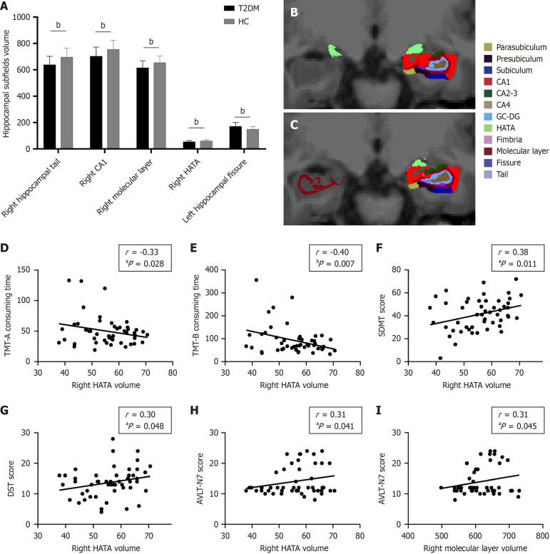
Methods: Sixty patients with T2DM and 32 healthy controls were recruited. All participants underwent a 3.0 T magnetic resonance scan and a series of clinical assessments. Hippocampal subfield volumes were determined using FreeSurfer 7.4.1. A two-sample t-test was used to evaluate group differences. Partial correlation analysis was performed to assess the relationship between hippocampal subregion volumes and cognitive function. aP < 0.05 was considered statistically significant.
Results: Compared with controls, the volume of right hippocampus-amygdala transition area (t = -3.053, P = 0.003) in patients with T2DM was significantly reduced, which was negatively correlated with the required time of the Trail Making Test (TMT)-A (r = -0.331, P = 0.028) and TMT-B (r = -0.402, P = 0.007) and positively correlated with the scores of Symbol Digit Modalities Test (r = 0.381, P = 0.011), Auditory Verbal Learning Test (AVLT)-N7 (r = 0.309, P = 0.041), and Digital Span Test (r = 0.300, P = 0.048). The volume of the right molecular layer (t = -2.998, P = 0.004) was also significantly reduced, which was positively associated with the scores of AVLT-N7 (r = 0.311, P = 0.045). In addition, the left hippocampal fissure volume (t = 3.617, P = 0.002) was significantly increased in patients with T2DM.
Conclusion: Declines in cognitive performance, especially memory and executive function, are linked to changes in the volumes of the right hippocampus-amygdala transition area and right molecular layer in patients with T2DM.
期刊介绍:
The WJD is a high-quality, peer reviewed, open-access journal. The primary task of WJD is to rapidly publish high-quality original articles, reviews, editorials, and case reports in the field of diabetes. In order to promote productive academic communication, the peer review process for the WJD is transparent; to this end, all published manuscripts are accompanied by the anonymized reviewers’ comments as well as the authors’ responses. The primary aims of the WJD are to improve diagnostic, therapeutic and preventive modalities and the skills of clinicians and to guide clinical practice in diabetes. Scope: Diabetes Complications, Experimental Diabetes Mellitus, Type 1 Diabetes Mellitus, Type 2 Diabetes Mellitus, Diabetes, Gestational, Diabetic Angiopathies, Diabetic Cardiomyopathies, Diabetic Coma, Diabetic Ketoacidosis, Diabetic Nephropathies, Diabetic Neuropathies, Donohue Syndrome, Fetal Macrosomia, and Prediabetic State.
 求助内容:
求助内容: 应助结果提醒方式:


