Jing Huang, Yating Li, Ming Zhang, Tiancheng Wu, Yuanzhen Zhang, Hui Wang
{"title":"产前不同剂量、阶段和疗程的阿莫西林暴露对子代卵巢发育的影响。","authors":"Jing Huang, Yating Li, Ming Zhang, Tiancheng Wu, Yuanzhen Zhang, Hui Wang","doi":"10.1186/s10020-025-01322-2","DOIUrl":null,"url":null,"abstract":"<p><strong>Background: </strong>Amoxicillin, a commonly used broad-spectrum penicillin antibiotic in pregnancy, has sparked controversy regarding its impact on fetal growth and development. There remains a lack of systematic research on the specific influence of prenatal amoxicillin exposure (PAmE) on the ovarian development of the offspring, as well as the precise \" toxicity windows \".</p><p><strong>Methods: </strong>We established PAmE mouse models at different stages [(gestational day, GD) 10-12, GD13-15 or GD16-18], doses (75, 150 or 300 mg/kg·d), and courses (single/multiple courses). On GD18, fetal serum and ovaries were collected to assess changes in serum estradiol levels and evaluate ovarian morphology, pregranulosa cell function, and oocyte-related parameters.</p><p><strong>Results: </strong>PAmE led to pathological damage in fetal mouse ovaries, characterized by disrupted germ cell cysts and reduced the number of germ cells. Cell proliferation was enhanced while apoptosis was reduced. Moreover, PAmE upregulated the expression of pregranulosa cell steroid synthesis-related genes (e.g., Sf1, Star, P450scc) in the fetal ovaries, particularly in the high-dose groups at all gestational stages. The expression of the oocyte marker gene Figlα increased in all PAmE groups, while follicle development-related genes (Nobox and Bmp15) were downregulated, particularly during early to mid-pregnancy and in the single-course exposure groups. Further investigation revealed that PAmE enhanced IGF1 expression in fetal ovaries and inhibited the Pten-Akt-Foxo3a signaling pathway.</p><p><strong>Conclusions: </strong>Amoxicillin exhibits ovarian developmental toxicity, influencing fetal ovarian cell proliferation, apoptosis, pregranulosa cell estrogen synthesis, oocyte numbers, and follicle assembly. This study provides evidence guiding the rational use of amoxicillin in pregnancy and assessing potential ovarian development risks.</p>","PeriodicalId":18813,"journal":{"name":"Molecular Medicine","volume":"31 1","pages":"261"},"PeriodicalIF":6.4000,"publicationDate":"2025-07-22","publicationTypes":"Journal Article","fieldsOfStudy":null,"isOpenAccess":false,"openAccessPdf":"https://www.ncbi.nlm.nih.gov/pmc/articles/PMC12285120/pdf/","citationCount":"0","resultStr":"{\"title\":\"The impact of prenatal amoxicillin exposure at different doses, stages, and courses on offspring ovarian development.\",\"authors\":\"Jing Huang, Yating Li, Ming Zhang, Tiancheng Wu, Yuanzhen Zhang, Hui Wang\",\"doi\":\"10.1186/s10020-025-01322-2\",\"DOIUrl\":null,\"url\":null,\"abstract\":\"<p><strong>Background: </strong>Amoxicillin, a commonly used broad-spectrum penicillin antibiotic in pregnancy, has sparked controversy regarding its impact on fetal growth and development. There remains a lack of systematic research on the specific influence of prenatal amoxicillin exposure (PAmE) on the ovarian development of the offspring, as well as the precise \\\" toxicity windows \\\".</p><p><strong>Methods: </strong>We established PAmE mouse models at different stages [(gestational day, GD) 10-12, GD13-15 or GD16-18], doses (75, 150 or 300 mg/kg·d), and courses (single/multiple courses). On GD18, fetal serum and ovaries were collected to assess changes in serum estradiol levels and evaluate ovarian morphology, pregranulosa cell function, and oocyte-related parameters.</p><p><strong>Results: </strong>PAmE led to pathological damage in fetal mouse ovaries, characterized by disrupted germ cell cysts and reduced the number of germ cells. Cell proliferation was enhanced while apoptosis was reduced. Moreover, PAmE upregulated the expression of pregranulosa cell steroid synthesis-related genes (e.g., Sf1, Star, P450scc) in the fetal ovaries, particularly in the high-dose groups at all gestational stages. The expression of the oocyte marker gene Figlα increased in all PAmE groups, while follicle development-related genes (Nobox and Bmp15) were downregulated, particularly during early to mid-pregnancy and in the single-course exposure groups. Further investigation revealed that PAmE enhanced IGF1 expression in fetal ovaries and inhibited the Pten-Akt-Foxo3a signaling pathway.</p><p><strong>Conclusions: </strong>Amoxicillin exhibits ovarian developmental toxicity, influencing fetal ovarian cell proliferation, apoptosis, pregranulosa cell estrogen synthesis, oocyte numbers, and follicle assembly. This study provides evidence guiding the rational use of amoxicillin in pregnancy and assessing potential ovarian development risks.</p>\",\"PeriodicalId\":18813,\"journal\":{\"name\":\"Molecular Medicine\",\"volume\":\"31 1\",\"pages\":\"261\"},\"PeriodicalIF\":6.4000,\"publicationDate\":\"2025-07-22\",\"publicationTypes\":\"Journal Article\",\"fieldsOfStudy\":null,\"isOpenAccess\":false,\"openAccessPdf\":\"https://www.ncbi.nlm.nih.gov/pmc/articles/PMC12285120/pdf/\",\"citationCount\":\"0\",\"resultStr\":null,\"platform\":\"Semanticscholar\",\"paperid\":null,\"PeriodicalName\":\"Molecular Medicine\",\"FirstCategoryId\":\"3\",\"ListUrlMain\":\"https://doi.org/10.1186/s10020-025-01322-2\",\"RegionNum\":2,\"RegionCategory\":\"医学\",\"ArticlePicture\":[],\"TitleCN\":null,\"AbstractTextCN\":null,\"PMCID\":null,\"EPubDate\":\"\",\"PubModel\":\"\",\"JCR\":\"Q1\",\"JCRName\":\"BIOCHEMISTRY & MOLECULAR BIOLOGY\",\"Score\":null,\"Total\":0}","platform":"Semanticscholar","paperid":null,"PeriodicalName":"Molecular Medicine","FirstCategoryId":"3","ListUrlMain":"https://doi.org/10.1186/s10020-025-01322-2","RegionNum":2,"RegionCategory":"医学","ArticlePicture":[],"TitleCN":null,"AbstractTextCN":null,"PMCID":null,"EPubDate":"","PubModel":"","JCR":"Q1","JCRName":"BIOCHEMISTRY & MOLECULAR BIOLOGY","Score":null,"Total":0}
The impact of prenatal amoxicillin exposure at different doses, stages, and courses on offspring ovarian development.
Background: Amoxicillin, a commonly used broad-spectrum penicillin antibiotic in pregnancy, has sparked controversy regarding its impact on fetal growth and development. There remains a lack of systematic research on the specific influence of prenatal amoxicillin exposure (PAmE) on the ovarian development of the offspring, as well as the precise " toxicity windows ".
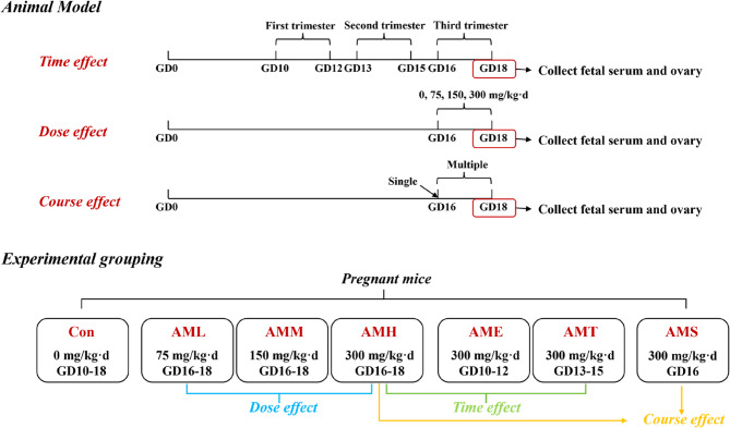
Methods: We established PAmE mouse models at different stages [(gestational day, GD) 10-12, GD13-15 or GD16-18], doses (75, 150 or 300 mg/kg·d), and courses (single/multiple courses). On GD18, fetal serum and ovaries were collected to assess changes in serum estradiol levels and evaluate ovarian morphology, pregranulosa cell function, and oocyte-related parameters.
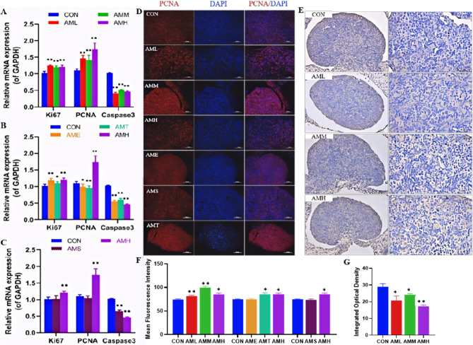
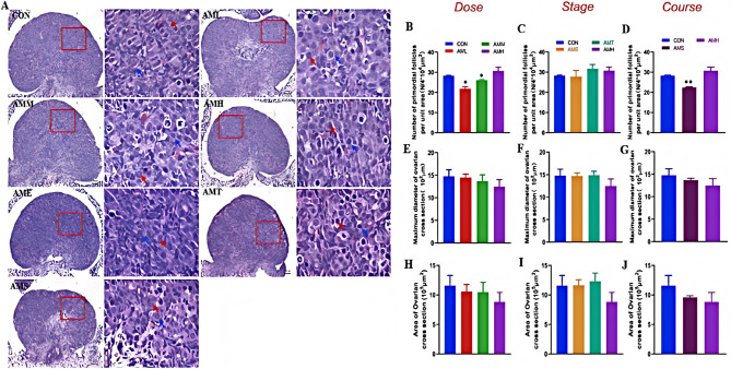
Results: PAmE led to pathological damage in fetal mouse ovaries, characterized by disrupted germ cell cysts and reduced the number of germ cells. Cell proliferation was enhanced while apoptosis was reduced. Moreover, PAmE upregulated the expression of pregranulosa cell steroid synthesis-related genes (e.g., Sf1, Star, P450scc) in the fetal ovaries, particularly in the high-dose groups at all gestational stages. The expression of the oocyte marker gene Figlα increased in all PAmE groups, while follicle development-related genes (Nobox and Bmp15) were downregulated, particularly during early to mid-pregnancy and in the single-course exposure groups. Further investigation revealed that PAmE enhanced IGF1 expression in fetal ovaries and inhibited the Pten-Akt-Foxo3a signaling pathway.
Conclusions: Amoxicillin exhibits ovarian developmental toxicity, influencing fetal ovarian cell proliferation, apoptosis, pregranulosa cell estrogen synthesis, oocyte numbers, and follicle assembly. This study provides evidence guiding the rational use of amoxicillin in pregnancy and assessing potential ovarian development risks.
期刊介绍:
Molecular Medicine is an open access journal that focuses on publishing recent findings related to disease pathogenesis at the molecular or physiological level. These insights can potentially contribute to the development of specific tools for disease diagnosis, treatment, or prevention. The journal considers manuscripts that present material pertinent to the genetic, molecular, or cellular underpinnings of critical physiological or disease processes. Submissions to Molecular Medicine are expected to elucidate the broader implications of the research findings for human disease and medicine in a manner that is accessible to a wide audience.
 求助内容:
求助内容: 应助结果提醒方式:


