Juan Zhang, Hongmei Luo, Yinqiao Li, Yayuan Feng, Xingpeng Pan, Beilei Ouyang, Guihong Nian, Ningyang Jia, Yonggang Li
{"title":"Gd-BOPTA增强MRI对无微血管侵袭的孤立性肝癌的预后价值。","authors":"Juan Zhang, Hongmei Luo, Yinqiao Li, Yayuan Feng, Xingpeng Pan, Beilei Ouyang, Guihong Nian, Ningyang Jia, Yonggang Li","doi":"10.2147/JHC.S530701","DOIUrl":null,"url":null,"abstract":"<p><strong>Objective: </strong>This study aims to evaluate the prognostic predictive efficacy of Gadobenate dimeglumine (Gd-BOPTA)-enhanced magnetic resonance imaging (MRI) in patients with solitary hepatocellular carcinoma (HCC) without microvascular invasion (MVI) and to investigate the potential clinical and imaging parameters for stratifying the risk of recurrence following hepatectomy.</p><p><strong>Methods: </strong>This retrospective study included 134 patients with histopathologically confirmed solitary HCC without microvascular invasion (MVI) from two hospital districts, which divided into the training cohort and validation cohort. MRI features were independently assessed by two radiologists. Univariate and multivariate Cox regression analyses were conducted to identify independent risk factors associated with recurrence-free survival (RFS). A nomogram was developed based on these factors, and its performance was validated in the validation cohort. RFS was analyzed using Kaplan-Meier curves and the Log rank test.</p><p><strong>Results: </strong>The median RFS for the 134 patients was 45.7 months, with 41.8% of patients experiencing tumor recurrence after hepatectomy. Univariate Cox regression analysis identified hepatitis Be antigen (HBeAg) positivity, tumor size, tumor growth subtype, non-peripheral washout, nodule-in-nodule architecture, mosaic architecture, and intratumoral arteries as significant risk factors for RFS. Multivariate Cox regression analysis revealed that HBeAg positive, tumor growth subtype, non-peripheral washout, mosaic architecture, and internal arteries were independent prognostic factors for RFS in patients with solitary HCC without MVI. The nomogram based on these variables demonstrated good predictive accuracy, with concordance indices (C-index) of 0.740 and 0.701 in the training and validation cohorts, respectively. Additionally, patients in the high-risk group exhibited significantly lower RFS compared to those in the low-risk group.</p><p><strong>Conclusion: </strong>A model incorporating Gd-BOPTA-enhanced MRI and clinical features can effectively predict RFS in solitary HCC patients without MVI and assist in risk stratification for recurrence after hepatectomy.</p>","PeriodicalId":15906,"journal":{"name":"Journal of Hepatocellular Carcinoma","volume":"12 ","pages":"1471-1482"},"PeriodicalIF":3.4000,"publicationDate":"2025-07-18","publicationTypes":"Journal Article","fieldsOfStudy":null,"isOpenAccess":false,"openAccessPdf":"https://www.ncbi.nlm.nih.gov/pmc/articles/PMC12282532/pdf/","citationCount":"0","resultStr":"{\"title\":\"Prognostic Value of Gd-BOPTA Enhanced MRI in Solitary Resected Hepatocellular Carcinoma Without Microvascular Invasion.\",\"authors\":\"Juan Zhang, Hongmei Luo, Yinqiao Li, Yayuan Feng, Xingpeng Pan, Beilei Ouyang, Guihong Nian, Ningyang Jia, Yonggang Li\",\"doi\":\"10.2147/JHC.S530701\",\"DOIUrl\":null,\"url\":null,\"abstract\":\"<p><strong>Objective: </strong>This study aims to evaluate the prognostic predictive efficacy of Gadobenate dimeglumine (Gd-BOPTA)-enhanced magnetic resonance imaging (MRI) in patients with solitary hepatocellular carcinoma (HCC) without microvascular invasion (MVI) and to investigate the potential clinical and imaging parameters for stratifying the risk of recurrence following hepatectomy.</p><p><strong>Methods: </strong>This retrospective study included 134 patients with histopathologically confirmed solitary HCC without microvascular invasion (MVI) from two hospital districts, which divided into the training cohort and validation cohort. MRI features were independently assessed by two radiologists. Univariate and multivariate Cox regression analyses were conducted to identify independent risk factors associated with recurrence-free survival (RFS). A nomogram was developed based on these factors, and its performance was validated in the validation cohort. RFS was analyzed using Kaplan-Meier curves and the Log rank test.</p><p><strong>Results: </strong>The median RFS for the 134 patients was 45.7 months, with 41.8% of patients experiencing tumor recurrence after hepatectomy. Univariate Cox regression analysis identified hepatitis Be antigen (HBeAg) positivity, tumor size, tumor growth subtype, non-peripheral washout, nodule-in-nodule architecture, mosaic architecture, and intratumoral arteries as significant risk factors for RFS. Multivariate Cox regression analysis revealed that HBeAg positive, tumor growth subtype, non-peripheral washout, mosaic architecture, and internal arteries were independent prognostic factors for RFS in patients with solitary HCC without MVI. The nomogram based on these variables demonstrated good predictive accuracy, with concordance indices (C-index) of 0.740 and 0.701 in the training and validation cohorts, respectively. Additionally, patients in the high-risk group exhibited significantly lower RFS compared to those in the low-risk group.</p><p><strong>Conclusion: </strong>A model incorporating Gd-BOPTA-enhanced MRI and clinical features can effectively predict RFS in solitary HCC patients without MVI and assist in risk stratification for recurrence after hepatectomy.</p>\",\"PeriodicalId\":15906,\"journal\":{\"name\":\"Journal of Hepatocellular Carcinoma\",\"volume\":\"12 \",\"pages\":\"1471-1482\"},\"PeriodicalIF\":3.4000,\"publicationDate\":\"2025-07-18\",\"publicationTypes\":\"Journal Article\",\"fieldsOfStudy\":null,\"isOpenAccess\":false,\"openAccessPdf\":\"https://www.ncbi.nlm.nih.gov/pmc/articles/PMC12282532/pdf/\",\"citationCount\":\"0\",\"resultStr\":null,\"platform\":\"Semanticscholar\",\"paperid\":null,\"PeriodicalName\":\"Journal of Hepatocellular Carcinoma\",\"FirstCategoryId\":\"3\",\"ListUrlMain\":\"https://doi.org/10.2147/JHC.S530701\",\"RegionNum\":3,\"RegionCategory\":\"医学\",\"ArticlePicture\":[],\"TitleCN\":null,\"AbstractTextCN\":null,\"PMCID\":null,\"EPubDate\":\"2025/1/1 0:00:00\",\"PubModel\":\"eCollection\",\"JCR\":\"Q2\",\"JCRName\":\"ONCOLOGY\",\"Score\":null,\"Total\":0}","platform":"Semanticscholar","paperid":null,"PeriodicalName":"Journal of Hepatocellular Carcinoma","FirstCategoryId":"3","ListUrlMain":"https://doi.org/10.2147/JHC.S530701","RegionNum":3,"RegionCategory":"医学","ArticlePicture":[],"TitleCN":null,"AbstractTextCN":null,"PMCID":null,"EPubDate":"2025/1/1 0:00:00","PubModel":"eCollection","JCR":"Q2","JCRName":"ONCOLOGY","Score":null,"Total":0}
Prognostic Value of Gd-BOPTA Enhanced MRI in Solitary Resected Hepatocellular Carcinoma Without Microvascular Invasion.
Objective: This study aims to evaluate the prognostic predictive efficacy of Gadobenate dimeglumine (Gd-BOPTA)-enhanced magnetic resonance imaging (MRI) in patients with solitary hepatocellular carcinoma (HCC) without microvascular invasion (MVI) and to investigate the potential clinical and imaging parameters for stratifying the risk of recurrence following hepatectomy.
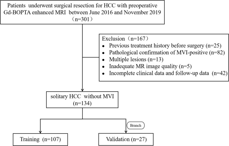
Methods: This retrospective study included 134 patients with histopathologically confirmed solitary HCC without microvascular invasion (MVI) from two hospital districts, which divided into the training cohort and validation cohort. MRI features were independently assessed by two radiologists. Univariate and multivariate Cox regression analyses were conducted to identify independent risk factors associated with recurrence-free survival (RFS). A nomogram was developed based on these factors, and its performance was validated in the validation cohort. RFS was analyzed using Kaplan-Meier curves and the Log rank test.
Results: The median RFS for the 134 patients was 45.7 months, with 41.8% of patients experiencing tumor recurrence after hepatectomy. Univariate Cox regression analysis identified hepatitis Be antigen (HBeAg) positivity, tumor size, tumor growth subtype, non-peripheral washout, nodule-in-nodule architecture, mosaic architecture, and intratumoral arteries as significant risk factors for RFS. Multivariate Cox regression analysis revealed that HBeAg positive, tumor growth subtype, non-peripheral washout, mosaic architecture, and internal arteries were independent prognostic factors for RFS in patients with solitary HCC without MVI. The nomogram based on these variables demonstrated good predictive accuracy, with concordance indices (C-index) of 0.740 and 0.701 in the training and validation cohorts, respectively. Additionally, patients in the high-risk group exhibited significantly lower RFS compared to those in the low-risk group.
Conclusion: A model incorporating Gd-BOPTA-enhanced MRI and clinical features can effectively predict RFS in solitary HCC patients without MVI and assist in risk stratification for recurrence after hepatectomy.
 求助内容:
求助内容: 应助结果提醒方式:


