Julia G Mannheim, Wenhong Lan, Maurizio Conti, Franziska Siedler, Marcel A Krueger, Kristina Herfert, Christian la Fougère, Fabian P Schmidt
{"title":"应用临床全身PET/CT系统进行小动物体内成像的可行性。","authors":"Julia G Mannheim, Wenhong Lan, Maurizio Conti, Franziska Siedler, Marcel A Krueger, Kristina Herfert, Christian la Fougère, Fabian P Schmidt","doi":"10.1186/s40658-025-00782-z","DOIUrl":null,"url":null,"abstract":"<p><strong>Background: </strong>Clinical PET scanners have long been explored for preclinical imaging, but limited spatial resolution and sensitivity have restricted their use for preclinical studies. The recent availability of total-body (TB) PET/CT scanners with extended axial fields of view (FOVs) has largely overcome sensitivity limitations, enabling potential new opportunities for small-animal imaging. This study evaluated the feasibility and performance of the Biograph Vision Quadra TB-PET/CT for rodent imaging compared to the dedicated small-animal PET scanner Inveon DPET.</p><p><strong>Material and methods: </strong>Recovery coefficients (RC), image noise, and optimum image reconstruction parameters were assessed using the preclinical NEMA NU 4-2008 image quality phantom and a sub-cohort of three anesthetized mice as a proof-of-concept demonstrating the feasibility of the setup. In vivo quantification accuracy was evaluated by scanning nine frozen mice simultaneously in three different arrangements with the Quadra compared with individual scans at the Inveon. To ensure comparability, all mice were snap-frozen after 1 h uptake of [<sup>1</sup>⁸F]FDG, scanned sequentially and individually at the Inveon (90 min p.i.), and subsequently scanned at the Quadra with decay-corrected acquisition times. SUV<sub>mean</sub> and SUV<sub>max</sub> values were determined for liver, muscle and brain regions on both systems. To evaluate potential position-dependent effects within the extended axial FOV, a single frozen mouse was scanned at multiple positions.</p><p><strong>Results: </strong>Phantom rods ≥ 2 mm could be resolved with the Quadra, showing a comparable RC for larger structures, e.g. for the 5 mm rod of 1.17 compared to 1.09 (Inveon) when using point-spread-function modeling, whilst having lower noise of 5.1%SD vs 9.0%SD. No substantial position-dependent effects were detected in the phantom or single-mouse scan across the axial FOV. SUV<sub>mean</sub> values were consistent between both scanner across all investigated organs, with liver and muscle uptake remaining comparable for frame durations down to 5 s. SUV<sub>max</sub> values exhibited greater variability, with significant differences observed in muscle and brain regions.</p><p><strong>Conclusion: </strong>Despite the lower spatial resolution of the clinical TB-PET/CT scanner (~ 3-4 mm) compared to the dedicated preclinical scanner (~ 1.5 mm), robust SUV<sub>mean</sub> quantification was achievable. Together with successful in vivo imaging of anesthetized mice, these findings support the feasibility of using clinical TB-PET/CT for preclinical research, acknowledging spatial resolution as a limiting factor.</p>","PeriodicalId":11559,"journal":{"name":"EJNMMI Physics","volume":"12 1","pages":"71"},"PeriodicalIF":3.2000,"publicationDate":"2025-07-23","publicationTypes":"Journal Article","fieldsOfStudy":null,"isOpenAccess":false,"openAccessPdf":"https://www.ncbi.nlm.nih.gov/pmc/articles/PMC12286903/pdf/","citationCount":"0","resultStr":"{\"title\":\"Feasibility of in vivo small animal imaging using a clinical total-body PET/CT system.\",\"authors\":\"Julia G Mannheim, Wenhong Lan, Maurizio Conti, Franziska Siedler, Marcel A Krueger, Kristina Herfert, Christian la Fougère, Fabian P Schmidt\",\"doi\":\"10.1186/s40658-025-00782-z\",\"DOIUrl\":null,\"url\":null,\"abstract\":\"<p><strong>Background: </strong>Clinical PET scanners have long been explored for preclinical imaging, but limited spatial resolution and sensitivity have restricted their use for preclinical studies. The recent availability of total-body (TB) PET/CT scanners with extended axial fields of view (FOVs) has largely overcome sensitivity limitations, enabling potential new opportunities for small-animal imaging. This study evaluated the feasibility and performance of the Biograph Vision Quadra TB-PET/CT for rodent imaging compared to the dedicated small-animal PET scanner Inveon DPET.</p><p><strong>Material and methods: </strong>Recovery coefficients (RC), image noise, and optimum image reconstruction parameters were assessed using the preclinical NEMA NU 4-2008 image quality phantom and a sub-cohort of three anesthetized mice as a proof-of-concept demonstrating the feasibility of the setup. In vivo quantification accuracy was evaluated by scanning nine frozen mice simultaneously in three different arrangements with the Quadra compared with individual scans at the Inveon. To ensure comparability, all mice were snap-frozen after 1 h uptake of [<sup>1</sup>⁸F]FDG, scanned sequentially and individually at the Inveon (90 min p.i.), and subsequently scanned at the Quadra with decay-corrected acquisition times. SUV<sub>mean</sub> and SUV<sub>max</sub> values were determined for liver, muscle and brain regions on both systems. To evaluate potential position-dependent effects within the extended axial FOV, a single frozen mouse was scanned at multiple positions.</p><p><strong>Results: </strong>Phantom rods ≥ 2 mm could be resolved with the Quadra, showing a comparable RC for larger structures, e.g. for the 5 mm rod of 1.17 compared to 1.09 (Inveon) when using point-spread-function modeling, whilst having lower noise of 5.1%SD vs 9.0%SD. No substantial position-dependent effects were detected in the phantom or single-mouse scan across the axial FOV. SUV<sub>mean</sub> values were consistent between both scanner across all investigated organs, with liver and muscle uptake remaining comparable for frame durations down to 5 s. SUV<sub>max</sub> values exhibited greater variability, with significant differences observed in muscle and brain regions.</p><p><strong>Conclusion: </strong>Despite the lower spatial resolution of the clinical TB-PET/CT scanner (~ 3-4 mm) compared to the dedicated preclinical scanner (~ 1.5 mm), robust SUV<sub>mean</sub> quantification was achievable. Together with successful in vivo imaging of anesthetized mice, these findings support the feasibility of using clinical TB-PET/CT for preclinical research, acknowledging spatial resolution as a limiting factor.</p>\",\"PeriodicalId\":11559,\"journal\":{\"name\":\"EJNMMI Physics\",\"volume\":\"12 1\",\"pages\":\"71\"},\"PeriodicalIF\":3.2000,\"publicationDate\":\"2025-07-23\",\"publicationTypes\":\"Journal Article\",\"fieldsOfStudy\":null,\"isOpenAccess\":false,\"openAccessPdf\":\"https://www.ncbi.nlm.nih.gov/pmc/articles/PMC12286903/pdf/\",\"citationCount\":\"0\",\"resultStr\":null,\"platform\":\"Semanticscholar\",\"paperid\":null,\"PeriodicalName\":\"EJNMMI Physics\",\"FirstCategoryId\":\"3\",\"ListUrlMain\":\"https://doi.org/10.1186/s40658-025-00782-z\",\"RegionNum\":2,\"RegionCategory\":\"医学\",\"ArticlePicture\":[],\"TitleCN\":null,\"AbstractTextCN\":null,\"PMCID\":null,\"EPubDate\":\"\",\"PubModel\":\"\",\"JCR\":\"Q2\",\"JCRName\":\"RADIOLOGY, NUCLEAR MEDICINE & MEDICAL IMAGING\",\"Score\":null,\"Total\":0}","platform":"Semanticscholar","paperid":null,"PeriodicalName":"EJNMMI Physics","FirstCategoryId":"3","ListUrlMain":"https://doi.org/10.1186/s40658-025-00782-z","RegionNum":2,"RegionCategory":"医学","ArticlePicture":[],"TitleCN":null,"AbstractTextCN":null,"PMCID":null,"EPubDate":"","PubModel":"","JCR":"Q2","JCRName":"RADIOLOGY, NUCLEAR MEDICINE & MEDICAL IMAGING","Score":null,"Total":0}
Feasibility of in vivo small animal imaging using a clinical total-body PET/CT system.
Background: Clinical PET scanners have long been explored for preclinical imaging, but limited spatial resolution and sensitivity have restricted their use for preclinical studies. The recent availability of total-body (TB) PET/CT scanners with extended axial fields of view (FOVs) has largely overcome sensitivity limitations, enabling potential new opportunities for small-animal imaging. This study evaluated the feasibility and performance of the Biograph Vision Quadra TB-PET/CT for rodent imaging compared to the dedicated small-animal PET scanner Inveon DPET.
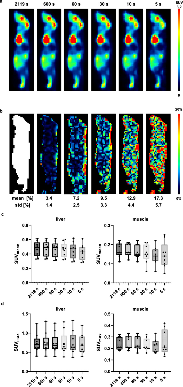
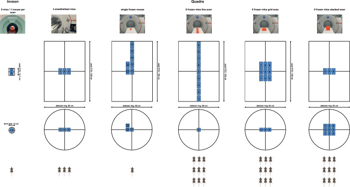
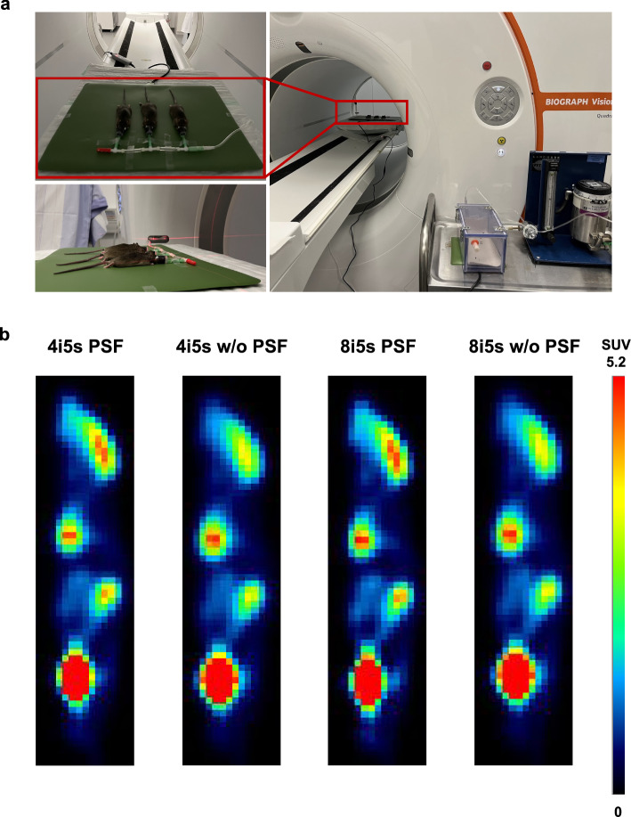
Material and methods: Recovery coefficients (RC), image noise, and optimum image reconstruction parameters were assessed using the preclinical NEMA NU 4-2008 image quality phantom and a sub-cohort of three anesthetized mice as a proof-of-concept demonstrating the feasibility of the setup. In vivo quantification accuracy was evaluated by scanning nine frozen mice simultaneously in three different arrangements with the Quadra compared with individual scans at the Inveon. To ensure comparability, all mice were snap-frozen after 1 h uptake of [1⁸F]FDG, scanned sequentially and individually at the Inveon (90 min p.i.), and subsequently scanned at the Quadra with decay-corrected acquisition times. SUVmean and SUVmax values were determined for liver, muscle and brain regions on both systems. To evaluate potential position-dependent effects within the extended axial FOV, a single frozen mouse was scanned at multiple positions.
Results: Phantom rods ≥ 2 mm could be resolved with the Quadra, showing a comparable RC for larger structures, e.g. for the 5 mm rod of 1.17 compared to 1.09 (Inveon) when using point-spread-function modeling, whilst having lower noise of 5.1%SD vs 9.0%SD. No substantial position-dependent effects were detected in the phantom or single-mouse scan across the axial FOV. SUVmean values were consistent between both scanner across all investigated organs, with liver and muscle uptake remaining comparable for frame durations down to 5 s. SUVmax values exhibited greater variability, with significant differences observed in muscle and brain regions.
Conclusion: Despite the lower spatial resolution of the clinical TB-PET/CT scanner (~ 3-4 mm) compared to the dedicated preclinical scanner (~ 1.5 mm), robust SUVmean quantification was achievable. Together with successful in vivo imaging of anesthetized mice, these findings support the feasibility of using clinical TB-PET/CT for preclinical research, acknowledging spatial resolution as a limiting factor.
期刊介绍:
EJNMMI Physics is an international platform for scientists, users and adopters of nuclear medicine with a particular interest in physics matters. As a companion journal to the European Journal of Nuclear Medicine and Molecular Imaging, this journal has a multi-disciplinary approach and welcomes original materials and studies with a focus on applied physics and mathematics as well as imaging systems engineering and prototyping in nuclear medicine. This includes physics-driven approaches or algorithms supported by physics that foster early clinical adoption of nuclear medicine imaging and therapy.

 求助内容:
求助内容: 应助结果提醒方式:


