Robin Hegering, Sindi Nexhipi, Theresa Suckert, Johannes Soltwedel, Elke Beyreuther, Mechthild Krause, Antje Dietrich, Armin Lühr
{"title":"局部脑质子辐照引起的小鼠反应性星形胶质细胞分布变化。","authors":"Robin Hegering, Sindi Nexhipi, Theresa Suckert, Johannes Soltwedel, Elke Beyreuther, Mechthild Krause, Antje Dietrich, Armin Lühr","doi":"10.2340/1651-226X.2025.44056","DOIUrl":null,"url":null,"abstract":"<p><strong>Background and purpose: </strong>After proton therapy of brain tumors, several studies have reported late image changes in follow-up magnetic resonance imaging, which result from blood-brain barrier (BBB) disruption. Astrocytes play a central role in the formation and maintenance of the BBB. To study the late response to partial-brain proton irradiation, preclinical mouse data were utilized to investigate the spatial distribution and dose dependence of reactive astrocytes.</p><p><strong>Material and methods: </strong>Previously, C57BL/6JRj mice were irradiated with protons targeting the right hippocampal region with single prescription doses of 45-85 Gy. After six months, mice were sacrificed and the excised brains axially cut into 3 µm thick slices and stained for glial fibrillary acidic protein (GFAP) to target astrocytes. Here, a workflow to segment the GFAP-positive area on slice images was established. The fraction of GFAP-positive area (GFAP+ fraction) was evaluated in the high-dose region in the right hemisphere and in the mirrored region in the left hemisphere. Dose distributions were simulated on pre-irradiation cone-beam computed tomography and co-registered to the histological slices.</p><p><strong>Results: </strong>For all irradiated mice, the GFAP+ fraction in the right hemisphere was significantly increased compared to the left hemisphere and to a sham-irradiated mouse with a highly symmetric GFAP distribution. The GFAP+ fraction in the right hemisphere increased approximately linearly with prescription dose. For comparable doses, the cerebral cortex showed lower GFAP+ fractions than the midbrain.</p><p><strong>Interpretation: </strong>GFAP upregulation correlated with dose level and distribution. In combination with other markers and timepoints, these findings contribute to a comprehensive understanding of cellular response.</p>","PeriodicalId":7110,"journal":{"name":"Acta Oncologica","volume":"64 ","pages":"902-908"},"PeriodicalIF":2.7000,"publicationDate":"2025-07-23","publicationTypes":"Journal Article","fieldsOfStudy":null,"isOpenAccess":false,"openAccessPdf":"https://www.ncbi.nlm.nih.gov/pmc/articles/PMC12305689/pdf/","citationCount":"0","resultStr":"{\"title\":\"Radiation-induced changes of reactive astrocyte distribution in mice as a late response to partial-brain proton irradiation.\",\"authors\":\"Robin Hegering, Sindi Nexhipi, Theresa Suckert, Johannes Soltwedel, Elke Beyreuther, Mechthild Krause, Antje Dietrich, Armin Lühr\",\"doi\":\"10.2340/1651-226X.2025.44056\",\"DOIUrl\":null,\"url\":null,\"abstract\":\"<p><strong>Background and purpose: </strong>After proton therapy of brain tumors, several studies have reported late image changes in follow-up magnetic resonance imaging, which result from blood-brain barrier (BBB) disruption. Astrocytes play a central role in the formation and maintenance of the BBB. To study the late response to partial-brain proton irradiation, preclinical mouse data were utilized to investigate the spatial distribution and dose dependence of reactive astrocytes.</p><p><strong>Material and methods: </strong>Previously, C57BL/6JRj mice were irradiated with protons targeting the right hippocampal region with single prescription doses of 45-85 Gy. After six months, mice were sacrificed and the excised brains axially cut into 3 µm thick slices and stained for glial fibrillary acidic protein (GFAP) to target astrocytes. Here, a workflow to segment the GFAP-positive area on slice images was established. The fraction of GFAP-positive area (GFAP+ fraction) was evaluated in the high-dose region in the right hemisphere and in the mirrored region in the left hemisphere. Dose distributions were simulated on pre-irradiation cone-beam computed tomography and co-registered to the histological slices.</p><p><strong>Results: </strong>For all irradiated mice, the GFAP+ fraction in the right hemisphere was significantly increased compared to the left hemisphere and to a sham-irradiated mouse with a highly symmetric GFAP distribution. The GFAP+ fraction in the right hemisphere increased approximately linearly with prescription dose. For comparable doses, the cerebral cortex showed lower GFAP+ fractions than the midbrain.</p><p><strong>Interpretation: </strong>GFAP upregulation correlated with dose level and distribution. In combination with other markers and timepoints, these findings contribute to a comprehensive understanding of cellular response.</p>\",\"PeriodicalId\":7110,\"journal\":{\"name\":\"Acta Oncologica\",\"volume\":\"64 \",\"pages\":\"902-908\"},\"PeriodicalIF\":2.7000,\"publicationDate\":\"2025-07-23\",\"publicationTypes\":\"Journal Article\",\"fieldsOfStudy\":null,\"isOpenAccess\":false,\"openAccessPdf\":\"https://www.ncbi.nlm.nih.gov/pmc/articles/PMC12305689/pdf/\",\"citationCount\":\"0\",\"resultStr\":null,\"platform\":\"Semanticscholar\",\"paperid\":null,\"PeriodicalName\":\"Acta Oncologica\",\"FirstCategoryId\":\"3\",\"ListUrlMain\":\"https://doi.org/10.2340/1651-226X.2025.44056\",\"RegionNum\":3,\"RegionCategory\":\"医学\",\"ArticlePicture\":[],\"TitleCN\":null,\"AbstractTextCN\":null,\"PMCID\":null,\"EPubDate\":\"\",\"PubModel\":\"\",\"JCR\":\"Q3\",\"JCRName\":\"ONCOLOGY\",\"Score\":null,\"Total\":0}","platform":"Semanticscholar","paperid":null,"PeriodicalName":"Acta Oncologica","FirstCategoryId":"3","ListUrlMain":"https://doi.org/10.2340/1651-226X.2025.44056","RegionNum":3,"RegionCategory":"医学","ArticlePicture":[],"TitleCN":null,"AbstractTextCN":null,"PMCID":null,"EPubDate":"","PubModel":"","JCR":"Q3","JCRName":"ONCOLOGY","Score":null,"Total":0}
Radiation-induced changes of reactive astrocyte distribution in mice as a late response to partial-brain proton irradiation.
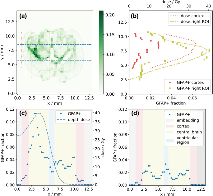
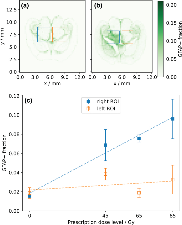
Background and purpose: After proton therapy of brain tumors, several studies have reported late image changes in follow-up magnetic resonance imaging, which result from blood-brain barrier (BBB) disruption. Astrocytes play a central role in the formation and maintenance of the BBB. To study the late response to partial-brain proton irradiation, preclinical mouse data were utilized to investigate the spatial distribution and dose dependence of reactive astrocytes.
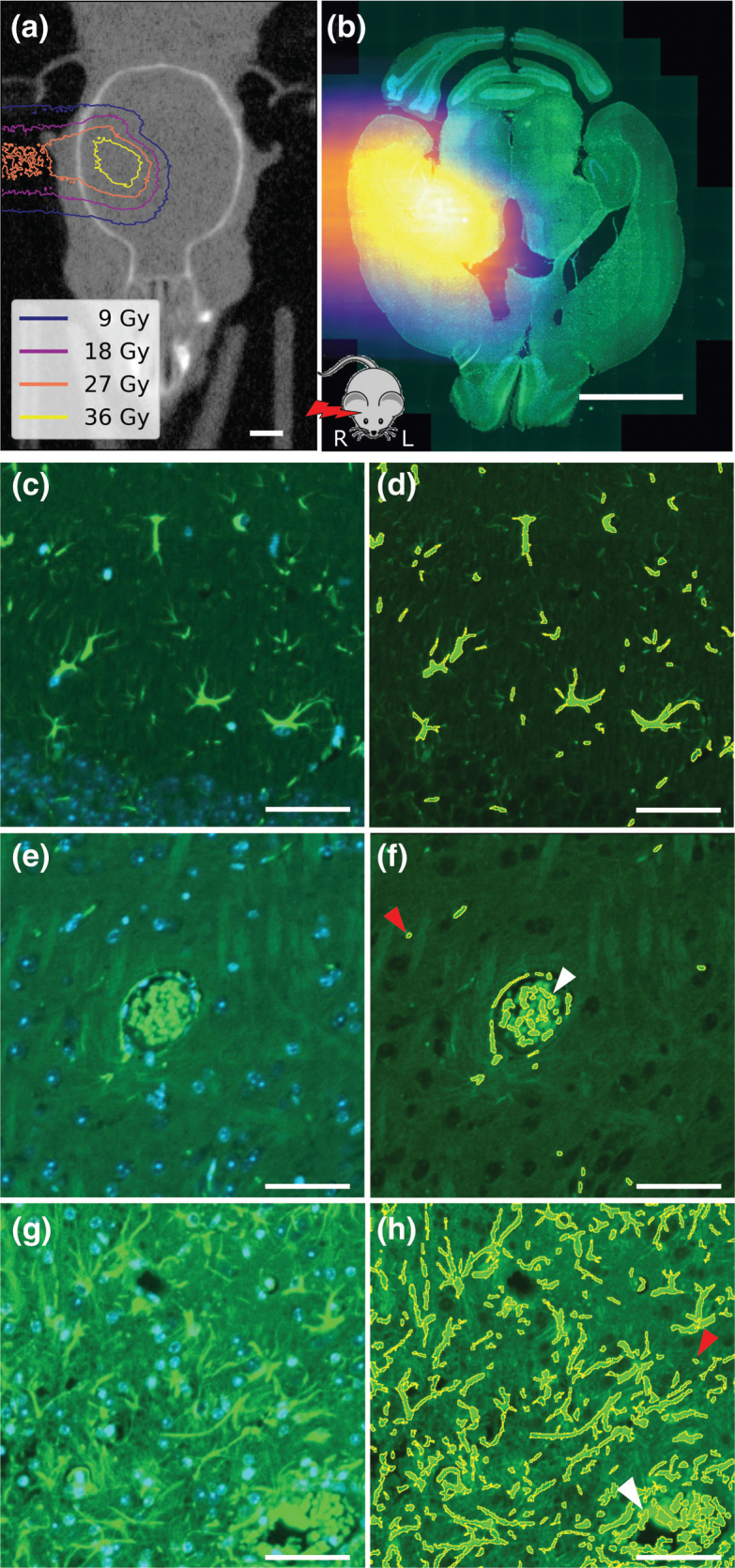
Material and methods: Previously, C57BL/6JRj mice were irradiated with protons targeting the right hippocampal region with single prescription doses of 45-85 Gy. After six months, mice were sacrificed and the excised brains axially cut into 3 µm thick slices and stained for glial fibrillary acidic protein (GFAP) to target astrocytes. Here, a workflow to segment the GFAP-positive area on slice images was established. The fraction of GFAP-positive area (GFAP+ fraction) was evaluated in the high-dose region in the right hemisphere and in the mirrored region in the left hemisphere. Dose distributions were simulated on pre-irradiation cone-beam computed tomography and co-registered to the histological slices.
Results: For all irradiated mice, the GFAP+ fraction in the right hemisphere was significantly increased compared to the left hemisphere and to a sham-irradiated mouse with a highly symmetric GFAP distribution. The GFAP+ fraction in the right hemisphere increased approximately linearly with prescription dose. For comparable doses, the cerebral cortex showed lower GFAP+ fractions than the midbrain.
Interpretation: GFAP upregulation correlated with dose level and distribution. In combination with other markers and timepoints, these findings contribute to a comprehensive understanding of cellular response.
期刊介绍:
Acta Oncologica is a journal for the clinical oncologist and accepts articles within all fields of clinical cancer research. Articles on tumour pathology, experimental oncology, radiobiology, cancer epidemiology and medical radio physics are also welcome, especially if they have a clinical aim or interest. Scientific articles on cancer nursing and psychological or social aspects of cancer are also welcomed. Extensive material may be published as Supplements, for which special conditions apply.
 求助内容:
求助内容: 应助结果提醒方式:


