{"title":"一种新型人工寰齿-齿状关节的设计及其经口咽入路关节成形术后的解剖学和放射学研究。","authors":"Shengguang Lei, Lingyong Jing, Xijiong Chen, Zhiwei Gan, Jianbin Zhong, Liming Yu, Yong Hu","doi":"10.1111/os.70088","DOIUrl":null,"url":null,"abstract":"<p><strong>Objective: </strong>For atlantoaxial joint disorders, traditional surgical approaches often presented challenges such as significant trauma and prolonged recovery. Therefore, it was crucial to explore safer and more effective surgical alternatives. The primary aim of this study was to investigate the anatomical safety and feasibility of artificial atlanto-odontoid joint (AAOJ) replacement via a transoral pharyngeal approach, through simulated surgical procedures and postoperative anatomical and radiological studies.</p><p><strong>Methods: </strong>The novel AAOJ replacement surgery was simulated on 18 fresh adult cadaveric head and neck specimens, and relevant anatomical parameters were measured. Postoperatively, the specimens underwent X-ray and CT scans, and software was used to measure the relevant parameters of the fixation screws. The spatial relationships between the atlantoaxial components, fixation screws, and critical anatomical structures were also examined. The comparison of parameters between the left and right sides was conducted using paired-sample t-tests.</p><p><strong>Results: </strong>The transoral pharyngeal approach provided adequate exposure, clear surgical visualization, and sufficient working space. Anatomical measurements showed that the width of the anterior arch bone window of the atlas was (13.8 ± 0.7) mm; the width of the vertebral body bone window of the axis was (11.0 ± 0.4) mm; the distance between the insertion points for the atlas screws was (28.2 ± 4.0) mm; the distance from the atlas insertion points to the lateral joint edge of the atlanto-axial joint was (5.2 ± 0.9) mm; the distance between the insertion points for the axis screws was (16.8 ± 1.6) mm; and the distance from the axis insertion points to the lateral joint edge of the atlanto-axial joint was (7.7 ± 0.9) mm. Radiological measurements showed that the screw trajectory length of the lateral mass screw in the atlas was (21.5 ± 2.8) mm, the outward insertion angle was (13.2 ± 2.5)°, and the caudal insertion angle was (3.5 ± 1.1)°; for the pedicle screw of the axis, the screw trajectory length was (29.8 ± 2.8) mm, the outward insertion angle was (20.7 ± 2.8)°, and the caudal insertion angle was (16.6 ± 2.7)°. The prosthesis was precisely fitted to the upper cervical spine, with adequate safety distances between the atlantoaxial components, fixation screws, and critical anatomical structures such as the foramen transversarium, vertebral artery groove, and spinal canal.</p><p><strong>Conclusions: </strong>The transoral pharyngeal approach for novel AAOJ replacement is anatomically safe and feasible.</p>","PeriodicalId":19566,"journal":{"name":"Orthopaedic Surgery","volume":" ","pages":"2670-2679"},"PeriodicalIF":2.1000,"publicationDate":"2025-09-01","publicationTypes":"Journal Article","fieldsOfStudy":null,"isOpenAccess":false,"openAccessPdf":"https://www.ncbi.nlm.nih.gov/pmc/articles/PMC12404860/pdf/","citationCount":"0","resultStr":"{\"title\":\"Design of a Novel Artificial Atlanto-Odontoid Joint and Its Anatomical and Radiological Studies Following Transoral Pharyngeal Approach Arthroplasty.\",\"authors\":\"Shengguang Lei, Lingyong Jing, Xijiong Chen, Zhiwei Gan, Jianbin Zhong, Liming Yu, Yong Hu\",\"doi\":\"10.1111/os.70088\",\"DOIUrl\":null,\"url\":null,\"abstract\":\"<p><strong>Objective: </strong>For atlantoaxial joint disorders, traditional surgical approaches often presented challenges such as significant trauma and prolonged recovery. Therefore, it was crucial to explore safer and more effective surgical alternatives. The primary aim of this study was to investigate the anatomical safety and feasibility of artificial atlanto-odontoid joint (AAOJ) replacement via a transoral pharyngeal approach, through simulated surgical procedures and postoperative anatomical and radiological studies.</p><p><strong>Methods: </strong>The novel AAOJ replacement surgery was simulated on 18 fresh adult cadaveric head and neck specimens, and relevant anatomical parameters were measured. Postoperatively, the specimens underwent X-ray and CT scans, and software was used to measure the relevant parameters of the fixation screws. The spatial relationships between the atlantoaxial components, fixation screws, and critical anatomical structures were also examined. The comparison of parameters between the left and right sides was conducted using paired-sample t-tests.</p><p><strong>Results: </strong>The transoral pharyngeal approach provided adequate exposure, clear surgical visualization, and sufficient working space. Anatomical measurements showed that the width of the anterior arch bone window of the atlas was (13.8 ± 0.7) mm; the width of the vertebral body bone window of the axis was (11.0 ± 0.4) mm; the distance between the insertion points for the atlas screws was (28.2 ± 4.0) mm; the distance from the atlas insertion points to the lateral joint edge of the atlanto-axial joint was (5.2 ± 0.9) mm; the distance between the insertion points for the axis screws was (16.8 ± 1.6) mm; and the distance from the axis insertion points to the lateral joint edge of the atlanto-axial joint was (7.7 ± 0.9) mm. Radiological measurements showed that the screw trajectory length of the lateral mass screw in the atlas was (21.5 ± 2.8) mm, the outward insertion angle was (13.2 ± 2.5)°, and the caudal insertion angle was (3.5 ± 1.1)°; for the pedicle screw of the axis, the screw trajectory length was (29.8 ± 2.8) mm, the outward insertion angle was (20.7 ± 2.8)°, and the caudal insertion angle was (16.6 ± 2.7)°. The prosthesis was precisely fitted to the upper cervical spine, with adequate safety distances between the atlantoaxial components, fixation screws, and critical anatomical structures such as the foramen transversarium, vertebral artery groove, and spinal canal.</p><p><strong>Conclusions: </strong>The transoral pharyngeal approach for novel AAOJ replacement is anatomically safe and feasible.</p>\",\"PeriodicalId\":19566,\"journal\":{\"name\":\"Orthopaedic Surgery\",\"volume\":\" \",\"pages\":\"2670-2679\"},\"PeriodicalIF\":2.1000,\"publicationDate\":\"2025-09-01\",\"publicationTypes\":\"Journal Article\",\"fieldsOfStudy\":null,\"isOpenAccess\":false,\"openAccessPdf\":\"https://www.ncbi.nlm.nih.gov/pmc/articles/PMC12404860/pdf/\",\"citationCount\":\"0\",\"resultStr\":null,\"platform\":\"Semanticscholar\",\"paperid\":null,\"PeriodicalName\":\"Orthopaedic Surgery\",\"FirstCategoryId\":\"3\",\"ListUrlMain\":\"https://doi.org/10.1111/os.70088\",\"RegionNum\":2,\"RegionCategory\":\"医学\",\"ArticlePicture\":[],\"TitleCN\":null,\"AbstractTextCN\":null,\"PMCID\":null,\"EPubDate\":\"2025/7/22 0:00:00\",\"PubModel\":\"Epub\",\"JCR\":\"Q2\",\"JCRName\":\"ORTHOPEDICS\",\"Score\":null,\"Total\":0}","platform":"Semanticscholar","paperid":null,"PeriodicalName":"Orthopaedic Surgery","FirstCategoryId":"3","ListUrlMain":"https://doi.org/10.1111/os.70088","RegionNum":2,"RegionCategory":"医学","ArticlePicture":[],"TitleCN":null,"AbstractTextCN":null,"PMCID":null,"EPubDate":"2025/7/22 0:00:00","PubModel":"Epub","JCR":"Q2","JCRName":"ORTHOPEDICS","Score":null,"Total":0}
Design of a Novel Artificial Atlanto-Odontoid Joint and Its Anatomical and Radiological Studies Following Transoral Pharyngeal Approach Arthroplasty.
Objective: For atlantoaxial joint disorders, traditional surgical approaches often presented challenges such as significant trauma and prolonged recovery. Therefore, it was crucial to explore safer and more effective surgical alternatives. The primary aim of this study was to investigate the anatomical safety and feasibility of artificial atlanto-odontoid joint (AAOJ) replacement via a transoral pharyngeal approach, through simulated surgical procedures and postoperative anatomical and radiological studies.
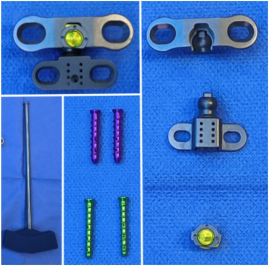
Methods: The novel AAOJ replacement surgery was simulated on 18 fresh adult cadaveric head and neck specimens, and relevant anatomical parameters were measured. Postoperatively, the specimens underwent X-ray and CT scans, and software was used to measure the relevant parameters of the fixation screws. The spatial relationships between the atlantoaxial components, fixation screws, and critical anatomical structures were also examined. The comparison of parameters between the left and right sides was conducted using paired-sample t-tests.
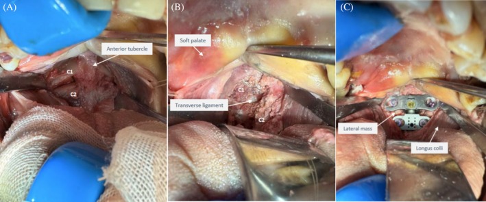
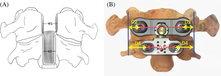
Results: The transoral pharyngeal approach provided adequate exposure, clear surgical visualization, and sufficient working space. Anatomical measurements showed that the width of the anterior arch bone window of the atlas was (13.8 ± 0.7) mm; the width of the vertebral body bone window of the axis was (11.0 ± 0.4) mm; the distance between the insertion points for the atlas screws was (28.2 ± 4.0) mm; the distance from the atlas insertion points to the lateral joint edge of the atlanto-axial joint was (5.2 ± 0.9) mm; the distance between the insertion points for the axis screws was (16.8 ± 1.6) mm; and the distance from the axis insertion points to the lateral joint edge of the atlanto-axial joint was (7.7 ± 0.9) mm. Radiological measurements showed that the screw trajectory length of the lateral mass screw in the atlas was (21.5 ± 2.8) mm, the outward insertion angle was (13.2 ± 2.5)°, and the caudal insertion angle was (3.5 ± 1.1)°; for the pedicle screw of the axis, the screw trajectory length was (29.8 ± 2.8) mm, the outward insertion angle was (20.7 ± 2.8)°, and the caudal insertion angle was (16.6 ± 2.7)°. The prosthesis was precisely fitted to the upper cervical spine, with adequate safety distances between the atlantoaxial components, fixation screws, and critical anatomical structures such as the foramen transversarium, vertebral artery groove, and spinal canal.
Conclusions: The transoral pharyngeal approach for novel AAOJ replacement is anatomically safe and feasible.
期刊介绍:
Orthopaedic Surgery (OS) is the official journal of the Chinese Orthopaedic Association, focusing on all aspects of orthopaedic technique and surgery.
The journal publishes peer-reviewed articles in the following categories: Original Articles, Clinical Articles, Review Articles, Guidelines, Editorials, Commentaries, Surgical Techniques, Case Reports and Meeting Reports.
 求助内容:
求助内容: 应助结果提醒方式:


