{"title":"气液界面培养人支气管上皮细胞在transwell膜上的分化及组织学分析","authors":"Asha Ashraf, Erica D. Bruce","doi":"10.1016/j.mex.2025.103492","DOIUrl":null,"url":null,"abstract":"<div><div>This protocol describes a reproducible and optimized workflow for the differentiation and histological analysis of primary normal human bronchial epithelial cells (NHBECs) cultured on Transwell membranes using air-liquid interface (ALI) culture. The method begins with expansion of early-passage NHBECs under submerged conditions, followed by seeding on collagen-coated Transwell inserts. Upon reaching confluence, cells are transitioned to ALI conditions using PneumaCult™-ALI maintenance medium for 21–28 days, promoting mucociliary differentiation. After differentiation, membranes are fixed, cryoprotected in graded sucrose solutions, embedded in Optimal Cutting Temperature (OCT) compound, and then cryosectioned. Sections are subsequently stained using hematoxylin and eosin (H&E) to assess epithelial morphology, or periodic acid–Schiff (PAS) staining to visualize polysaccharide-containing structures such as mucins. This protocol supports the detailed structural and histological evaluation of NHBEC differentiation on a Transwell model, which is valuable for studies of airway epithelial biology, mucociliary function, and inhalation toxicology.<ul><li><span>•</span><span><div>ALI differentiation of NHBECs on collagen-coated Transwell inserts.</div></span></li><li><span>•</span><span><div>Fixation, sucrose cryoprotection, and OCT embedding for cryosectioning.</div></span></li><li><span>•</span><span><div>H&E and PAS staining for visualization of epithelial cells and mucin.</div></span></li></ul></div></div>","PeriodicalId":18446,"journal":{"name":"MethodsX","volume":"15 ","pages":"Article 103492"},"PeriodicalIF":1.9000,"publicationDate":"2025-07-07","publicationTypes":"Journal Article","fieldsOfStudy":null,"isOpenAccess":false,"openAccessPdf":"","citationCount":"0","resultStr":"{\"title\":\"Differentiation and histological analysis of primary human bronchial epithelial cells cultured on transwell membranes using air-liquid interface culture\",\"authors\":\"Asha Ashraf, Erica D. Bruce\",\"doi\":\"10.1016/j.mex.2025.103492\",\"DOIUrl\":null,\"url\":null,\"abstract\":\"<div><div>This protocol describes a reproducible and optimized workflow for the differentiation and histological analysis of primary normal human bronchial epithelial cells (NHBECs) cultured on Transwell membranes using air-liquid interface (ALI) culture. The method begins with expansion of early-passage NHBECs under submerged conditions, followed by seeding on collagen-coated Transwell inserts. Upon reaching confluence, cells are transitioned to ALI conditions using PneumaCult™-ALI maintenance medium for 21–28 days, promoting mucociliary differentiation. After differentiation, membranes are fixed, cryoprotected in graded sucrose solutions, embedded in Optimal Cutting Temperature (OCT) compound, and then cryosectioned. Sections are subsequently stained using hematoxylin and eosin (H&E) to assess epithelial morphology, or periodic acid–Schiff (PAS) staining to visualize polysaccharide-containing structures such as mucins. This protocol supports the detailed structural and histological evaluation of NHBEC differentiation on a Transwell model, which is valuable for studies of airway epithelial biology, mucociliary function, and inhalation toxicology.<ul><li><span>•</span><span><div>ALI differentiation of NHBECs on collagen-coated Transwell inserts.</div></span></li><li><span>•</span><span><div>Fixation, sucrose cryoprotection, and OCT embedding for cryosectioning.</div></span></li><li><span>•</span><span><div>H&E and PAS staining for visualization of epithelial cells and mucin.</div></span></li></ul></div></div>\",\"PeriodicalId\":18446,\"journal\":{\"name\":\"MethodsX\",\"volume\":\"15 \",\"pages\":\"Article 103492\"},\"PeriodicalIF\":1.9000,\"publicationDate\":\"2025-07-07\",\"publicationTypes\":\"Journal Article\",\"fieldsOfStudy\":null,\"isOpenAccess\":false,\"openAccessPdf\":\"\",\"citationCount\":\"0\",\"resultStr\":null,\"platform\":\"Semanticscholar\",\"paperid\":null,\"PeriodicalName\":\"MethodsX\",\"FirstCategoryId\":\"1085\",\"ListUrlMain\":\"https://www.sciencedirect.com/science/article/pii/S2215016125003371\",\"RegionNum\":0,\"RegionCategory\":null,\"ArticlePicture\":[],\"TitleCN\":null,\"AbstractTextCN\":null,\"PMCID\":null,\"EPubDate\":\"\",\"PubModel\":\"\",\"JCR\":\"Q2\",\"JCRName\":\"MULTIDISCIPLINARY SCIENCES\",\"Score\":null,\"Total\":0}","platform":"Semanticscholar","paperid":null,"PeriodicalName":"MethodsX","FirstCategoryId":"1085","ListUrlMain":"https://www.sciencedirect.com/science/article/pii/S2215016125003371","RegionNum":0,"RegionCategory":null,"ArticlePicture":[],"TitleCN":null,"AbstractTextCN":null,"PMCID":null,"EPubDate":"","PubModel":"","JCR":"Q2","JCRName":"MULTIDISCIPLINARY SCIENCES","Score":null,"Total":0}
Differentiation and histological analysis of primary human bronchial epithelial cells cultured on transwell membranes using air-liquid interface culture
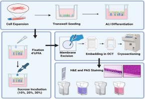
This protocol describes a reproducible and optimized workflow for the differentiation and histological analysis of primary normal human bronchial epithelial cells (NHBECs) cultured on Transwell membranes using air-liquid interface (ALI) culture. The method begins with expansion of early-passage NHBECs under submerged conditions, followed by seeding on collagen-coated Transwell inserts. Upon reaching confluence, cells are transitioned to ALI conditions using PneumaCult™-ALI maintenance medium for 21–28 days, promoting mucociliary differentiation. After differentiation, membranes are fixed, cryoprotected in graded sucrose solutions, embedded in Optimal Cutting Temperature (OCT) compound, and then cryosectioned. Sections are subsequently stained using hematoxylin and eosin (H&E) to assess epithelial morphology, or periodic acid–Schiff (PAS) staining to visualize polysaccharide-containing structures such as mucins. This protocol supports the detailed structural and histological evaluation of NHBEC differentiation on a Transwell model, which is valuable for studies of airway epithelial biology, mucociliary function, and inhalation toxicology.
•
ALI differentiation of NHBECs on collagen-coated Transwell inserts.
•
Fixation, sucrose cryoprotection, and OCT embedding for cryosectioning.
•
H&E and PAS staining for visualization of epithelial cells and mucin.