Ashish Kumar Jha, Umeshkumar Baburao Sherkhane, Nilendu C Purandare, Leonard Wee, Andre Dekker, Venkatesh Rangarajan
{"title":"正电子发射断层成像生物标志物和人工智能表征孤立性肺结节。","authors":"Ashish Kumar Jha, Umeshkumar Baburao Sherkhane, Nilendu C Purandare, Leonard Wee, Andre Dekker, Venkatesh Rangarajan","doi":"10.3389/fnume.2025.1611823","DOIUrl":null,"url":null,"abstract":"<p><strong>Background: </strong>The characterization of solitary pulmonary nodules (SPNs) as malignant or benign remains a diagnostic challenge using conventional imaging parameters. The literature suggests using combined Positron Emission Tomography (PET) and Computed Tomography (CT) to characterise a SPN. Radiomics and machine learning are other promising technologies which can be utilised to characterise the SPN.</p><p><strong>Purpose: </strong>This study explores the potential of PET radiomics signatures and machine learning algorithms to characterise the SPN.</p><p><strong>Methods: </strong>This retrospective study aimed to characterize solitary pulmonary nodules (SPNs) using PET radiomics. A total of 163 patients who underwent PET/CT imaging were included in this study. A total of 1,098 features were extracted from PET images using PyRadiomics. To optimize model performance two strategies i.e., (a) feature selection and (b) feature reduction techniques were employed, including hierarchical clustering, RFE in feature selection, and PCA in feature reduction. To address outcome class imbalance, the dataset was statistically resampled (SMOTE). A random forest models was developed using original training set (RF-Model-O & RF-PCA-Model-O) and balanced training dataset (RF-Model-B & RF-PCA-Model-B) and validated on the test datasets. Additionally, 5-fold cross-validation and bootstrap validation was also performed. The model's performance was assessed using various metrics, such as accuracy, AUC, precision, recall, and F1-score.</p><p><strong>Results: </strong>Of the 163 patients (aged 36-76 years, mean age 58 ± 7), 117 had malignant disease and 46 had granulomatous or benign conditions. In <b>Strategy (a),</b> five radiomic features were identified as optimal using hierarchical clustering and RFE. In <b>Strategy (b),</b> five principal components were deemed optimal using PCA. The model accuracy of RF-Model-O and RF-Model-B in the train-test validation, 5-fold cross-validation and bootstrap validation were found to be 0.8, 0.80 ± 0.07, 0.84 ± 1.11 and 0.8, 0.83 ± 0.10, 0.80 ± 0.07 in Strategy (a). Similarly, the model accuracy of RF-PCA-Model-O and RF-PCA-Model-B in the train-test validation, 5-fold cross-validation and bootstrap validation were found to be 0.84, 0.80 ± 0.07, 0.84 ± 07 and 0.74, 0.80 ± 0.08, 0.75 ± 0.08 in Strategy (b).</p><p><strong>Conclusion: </strong>The PET radiomics demonstrated excellent performance in characterizing SPNs as benign or malignant.</p>","PeriodicalId":73095,"journal":{"name":"Frontiers in nuclear medicine (Lausanne, Switzerland)","volume":"5 ","pages":"1611823"},"PeriodicalIF":1.4000,"publicationDate":"2025-07-04","publicationTypes":"Journal Article","fieldsOfStudy":null,"isOpenAccess":false,"openAccessPdf":"https://www.ncbi.nlm.nih.gov/pmc/articles/PMC12271206/pdf/","citationCount":"0","resultStr":"{\"title\":\"Positron emission tomography imaging biomarker and artificial intelligence for the characterization of solitary pulmonary nodule.\",\"authors\":\"Ashish Kumar Jha, Umeshkumar Baburao Sherkhane, Nilendu C Purandare, Leonard Wee, Andre Dekker, Venkatesh Rangarajan\",\"doi\":\"10.3389/fnume.2025.1611823\",\"DOIUrl\":null,\"url\":null,\"abstract\":\"<p><strong>Background: </strong>The characterization of solitary pulmonary nodules (SPNs) as malignant or benign remains a diagnostic challenge using conventional imaging parameters. The literature suggests using combined Positron Emission Tomography (PET) and Computed Tomography (CT) to characterise a SPN. Radiomics and machine learning are other promising technologies which can be utilised to characterise the SPN.</p><p><strong>Purpose: </strong>This study explores the potential of PET radiomics signatures and machine learning algorithms to characterise the SPN.</p><p><strong>Methods: </strong>This retrospective study aimed to characterize solitary pulmonary nodules (SPNs) using PET radiomics. A total of 163 patients who underwent PET/CT imaging were included in this study. A total of 1,098 features were extracted from PET images using PyRadiomics. To optimize model performance two strategies i.e., (a) feature selection and (b) feature reduction techniques were employed, including hierarchical clustering, RFE in feature selection, and PCA in feature reduction. To address outcome class imbalance, the dataset was statistically resampled (SMOTE). A random forest models was developed using original training set (RF-Model-O & RF-PCA-Model-O) and balanced training dataset (RF-Model-B & RF-PCA-Model-B) and validated on the test datasets. Additionally, 5-fold cross-validation and bootstrap validation was also performed. The model's performance was assessed using various metrics, such as accuracy, AUC, precision, recall, and F1-score.</p><p><strong>Results: </strong>Of the 163 patients (aged 36-76 years, mean age 58 ± 7), 117 had malignant disease and 46 had granulomatous or benign conditions. In <b>Strategy (a),</b> five radiomic features were identified as optimal using hierarchical clustering and RFE. In <b>Strategy (b),</b> five principal components were deemed optimal using PCA. The model accuracy of RF-Model-O and RF-Model-B in the train-test validation, 5-fold cross-validation and bootstrap validation were found to be 0.8, 0.80 ± 0.07, 0.84 ± 1.11 and 0.8, 0.83 ± 0.10, 0.80 ± 0.07 in Strategy (a). Similarly, the model accuracy of RF-PCA-Model-O and RF-PCA-Model-B in the train-test validation, 5-fold cross-validation and bootstrap validation were found to be 0.84, 0.80 ± 0.07, 0.84 ± 07 and 0.74, 0.80 ± 0.08, 0.75 ± 0.08 in Strategy (b).</p><p><strong>Conclusion: </strong>The PET radiomics demonstrated excellent performance in characterizing SPNs as benign or malignant.</p>\",\"PeriodicalId\":73095,\"journal\":{\"name\":\"Frontiers in nuclear medicine (Lausanne, Switzerland)\",\"volume\":\"5 \",\"pages\":\"1611823\"},\"PeriodicalIF\":1.4000,\"publicationDate\":\"2025-07-04\",\"publicationTypes\":\"Journal Article\",\"fieldsOfStudy\":null,\"isOpenAccess\":false,\"openAccessPdf\":\"https://www.ncbi.nlm.nih.gov/pmc/articles/PMC12271206/pdf/\",\"citationCount\":\"0\",\"resultStr\":null,\"platform\":\"Semanticscholar\",\"paperid\":null,\"PeriodicalName\":\"Frontiers in nuclear medicine (Lausanne, Switzerland)\",\"FirstCategoryId\":\"1085\",\"ListUrlMain\":\"https://doi.org/10.3389/fnume.2025.1611823\",\"RegionNum\":0,\"RegionCategory\":null,\"ArticlePicture\":[],\"TitleCN\":null,\"AbstractTextCN\":null,\"PMCID\":null,\"EPubDate\":\"2025/1/1 0:00:00\",\"PubModel\":\"eCollection\",\"JCR\":\"\",\"JCRName\":\"\",\"Score\":null,\"Total\":0}","platform":"Semanticscholar","paperid":null,"PeriodicalName":"Frontiers in nuclear medicine (Lausanne, Switzerland)","FirstCategoryId":"1085","ListUrlMain":"https://doi.org/10.3389/fnume.2025.1611823","RegionNum":0,"RegionCategory":null,"ArticlePicture":[],"TitleCN":null,"AbstractTextCN":null,"PMCID":null,"EPubDate":"2025/1/1 0:00:00","PubModel":"eCollection","JCR":"","JCRName":"","Score":null,"Total":0}
Positron emission tomography imaging biomarker and artificial intelligence for the characterization of solitary pulmonary nodule.
Background: The characterization of solitary pulmonary nodules (SPNs) as malignant or benign remains a diagnostic challenge using conventional imaging parameters. The literature suggests using combined Positron Emission Tomography (PET) and Computed Tomography (CT) to characterise a SPN. Radiomics and machine learning are other promising technologies which can be utilised to characterise the SPN.
Purpose: This study explores the potential of PET radiomics signatures and machine learning algorithms to characterise the SPN.
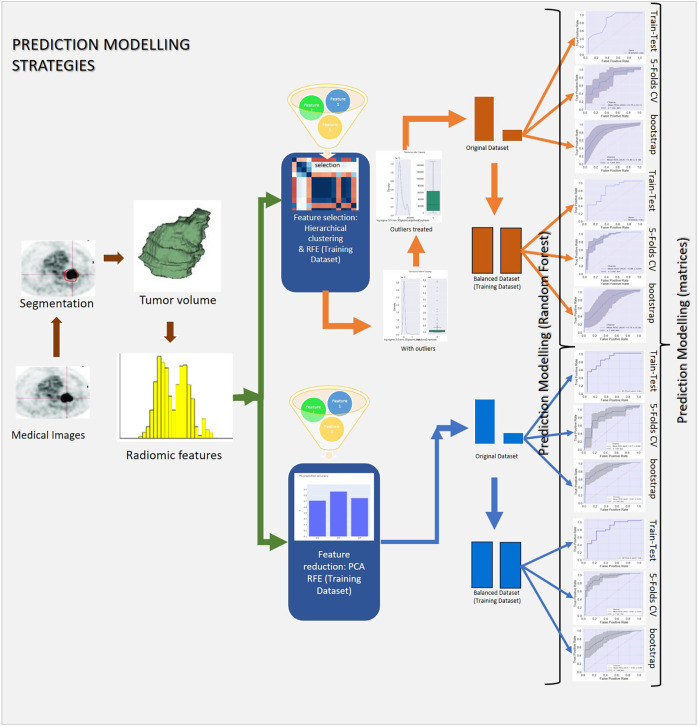
Methods: This retrospective study aimed to characterize solitary pulmonary nodules (SPNs) using PET radiomics. A total of 163 patients who underwent PET/CT imaging were included in this study. A total of 1,098 features were extracted from PET images using PyRadiomics. To optimize model performance two strategies i.e., (a) feature selection and (b) feature reduction techniques were employed, including hierarchical clustering, RFE in feature selection, and PCA in feature reduction. To address outcome class imbalance, the dataset was statistically resampled (SMOTE). A random forest models was developed using original training set (RF-Model-O & RF-PCA-Model-O) and balanced training dataset (RF-Model-B & RF-PCA-Model-B) and validated on the test datasets. Additionally, 5-fold cross-validation and bootstrap validation was also performed. The model's performance was assessed using various metrics, such as accuracy, AUC, precision, recall, and F1-score.
Results: Of the 163 patients (aged 36-76 years, mean age 58 ± 7), 117 had malignant disease and 46 had granulomatous or benign conditions. In Strategy (a), five radiomic features were identified as optimal using hierarchical clustering and RFE. In Strategy (b), five principal components were deemed optimal using PCA. The model accuracy of RF-Model-O and RF-Model-B in the train-test validation, 5-fold cross-validation and bootstrap validation were found to be 0.8, 0.80 ± 0.07, 0.84 ± 1.11 and 0.8, 0.83 ± 0.10, 0.80 ± 0.07 in Strategy (a). Similarly, the model accuracy of RF-PCA-Model-O and RF-PCA-Model-B in the train-test validation, 5-fold cross-validation and bootstrap validation were found to be 0.84, 0.80 ± 0.07, 0.84 ± 07 and 0.74, 0.80 ± 0.08, 0.75 ± 0.08 in Strategy (b).
Conclusion: The PET radiomics demonstrated excellent performance in characterizing SPNs as benign or malignant.


 求助内容:
求助内容: 应助结果提醒方式:


