Le Guo, Chantao Huang, Peng Hao, Ningyang Jia, Ling Zhang
{"title":"基于MRI的瘤周放射组学能否预测肝细胞癌合并胆管癌术前微血管侵袭状况?","authors":"Le Guo, Chantao Huang, Peng Hao, Ningyang Jia, Ling Zhang","doi":"10.2147/JHC.S515651","DOIUrl":null,"url":null,"abstract":"<p><strong>Objective: </strong>To investigate the role of MRI peritumoral imaging in predicting microvascular invasion (MVI) status in patients with combined hepatocellular carcinoma and cholangiocarcinoma (cHCC-CCA).</p><p><strong>Methods: </strong>Clinical and pathological data and MRI images of 118 patients with surgically resected and pathologically confirmed cHCC-CCA were retrospectively collected. The tumor in MRI images was segmented by ITK-SNAP software in three dimensions and extended 1 centimeter(cm) towards the tumor periphery. Then, the Python open-source platform was used for radiomics analysis. Mutual information and recursive elimination methods were used to select the optimal features. Clinical models and radiomics models were constructed based on six classifiers. The model's effectiveness was comprehensively evaluated using receiver operating characteristic (ROC), area under curve (AUC), and decision curve analysis (DCA), and the model results were output using Shapley Additive exPlans (SHAP).</p><p><strong>Results: </strong>The differences in HBeAg, capsule, target sign, and lymph node metastasis between MVI negative and positive groups were statistically significant (<i>p</i> < 0.05). Based on peritumoral, 1cm fusion model (in arterial phase) has an AUC of 0.940 (95% CI: 0.801-0.947) and 0.825 (95% CI: 0.633-0.917) in the training/testing set when identifying the MVI status of cHCC-CCA. The accuracy, sensitivity, and specificity in the testing set are 0.778, 0.800, and 0.726, respectively. The DCA shows that when the threshold is approximately 11.08%-66.47%, the net return of the fusion model is higher than that of the clinical and radiomics models under the same conditions.</p><p><strong>Conclusion: </strong>Radiomics with a 1cm extension around the tumor can improve the performance of machine-learning models in predicting MVI labels.</p>","PeriodicalId":15906,"journal":{"name":"Journal of Hepatocellular Carcinoma","volume":"12 ","pages":"1441-1452"},"PeriodicalIF":3.4000,"publicationDate":"2025-07-16","publicationTypes":"Journal Article","fieldsOfStudy":null,"isOpenAccess":false,"openAccessPdf":"https://www.ncbi.nlm.nih.gov/pmc/articles/PMC12276744/pdf/","citationCount":"0","resultStr":"{\"title\":\"Can Peritumoral Radiomics Based on MRI Predict the Microvascular Invasion Status of Combined Hepatocellular Carcinoma and Cholangiocarcinoma Before Surgery?\",\"authors\":\"Le Guo, Chantao Huang, Peng Hao, Ningyang Jia, Ling Zhang\",\"doi\":\"10.2147/JHC.S515651\",\"DOIUrl\":null,\"url\":null,\"abstract\":\"<p><strong>Objective: </strong>To investigate the role of MRI peritumoral imaging in predicting microvascular invasion (MVI) status in patients with combined hepatocellular carcinoma and cholangiocarcinoma (cHCC-CCA).</p><p><strong>Methods: </strong>Clinical and pathological data and MRI images of 118 patients with surgically resected and pathologically confirmed cHCC-CCA were retrospectively collected. The tumor in MRI images was segmented by ITK-SNAP software in three dimensions and extended 1 centimeter(cm) towards the tumor periphery. Then, the Python open-source platform was used for radiomics analysis. Mutual information and recursive elimination methods were used to select the optimal features. Clinical models and radiomics models were constructed based on six classifiers. The model's effectiveness was comprehensively evaluated using receiver operating characteristic (ROC), area under curve (AUC), and decision curve analysis (DCA), and the model results were output using Shapley Additive exPlans (SHAP).</p><p><strong>Results: </strong>The differences in HBeAg, capsule, target sign, and lymph node metastasis between MVI negative and positive groups were statistically significant (<i>p</i> < 0.05). Based on peritumoral, 1cm fusion model (in arterial phase) has an AUC of 0.940 (95% CI: 0.801-0.947) and 0.825 (95% CI: 0.633-0.917) in the training/testing set when identifying the MVI status of cHCC-CCA. The accuracy, sensitivity, and specificity in the testing set are 0.778, 0.800, and 0.726, respectively. The DCA shows that when the threshold is approximately 11.08%-66.47%, the net return of the fusion model is higher than that of the clinical and radiomics models under the same conditions.</p><p><strong>Conclusion: </strong>Radiomics with a 1cm extension around the tumor can improve the performance of machine-learning models in predicting MVI labels.</p>\",\"PeriodicalId\":15906,\"journal\":{\"name\":\"Journal of Hepatocellular Carcinoma\",\"volume\":\"12 \",\"pages\":\"1441-1452\"},\"PeriodicalIF\":3.4000,\"publicationDate\":\"2025-07-16\",\"publicationTypes\":\"Journal Article\",\"fieldsOfStudy\":null,\"isOpenAccess\":false,\"openAccessPdf\":\"https://www.ncbi.nlm.nih.gov/pmc/articles/PMC12276744/pdf/\",\"citationCount\":\"0\",\"resultStr\":null,\"platform\":\"Semanticscholar\",\"paperid\":null,\"PeriodicalName\":\"Journal of Hepatocellular Carcinoma\",\"FirstCategoryId\":\"3\",\"ListUrlMain\":\"https://doi.org/10.2147/JHC.S515651\",\"RegionNum\":3,\"RegionCategory\":\"医学\",\"ArticlePicture\":[],\"TitleCN\":null,\"AbstractTextCN\":null,\"PMCID\":null,\"EPubDate\":\"2025/1/1 0:00:00\",\"PubModel\":\"eCollection\",\"JCR\":\"Q2\",\"JCRName\":\"ONCOLOGY\",\"Score\":null,\"Total\":0}","platform":"Semanticscholar","paperid":null,"PeriodicalName":"Journal of Hepatocellular Carcinoma","FirstCategoryId":"3","ListUrlMain":"https://doi.org/10.2147/JHC.S515651","RegionNum":3,"RegionCategory":"医学","ArticlePicture":[],"TitleCN":null,"AbstractTextCN":null,"PMCID":null,"EPubDate":"2025/1/1 0:00:00","PubModel":"eCollection","JCR":"Q2","JCRName":"ONCOLOGY","Score":null,"Total":0}
Can Peritumoral Radiomics Based on MRI Predict the Microvascular Invasion Status of Combined Hepatocellular Carcinoma and Cholangiocarcinoma Before Surgery?
Objective: To investigate the role of MRI peritumoral imaging in predicting microvascular invasion (MVI) status in patients with combined hepatocellular carcinoma and cholangiocarcinoma (cHCC-CCA).
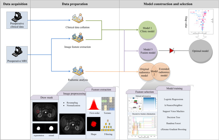
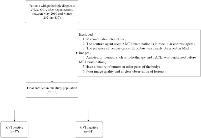
Methods: Clinical and pathological data and MRI images of 118 patients with surgically resected and pathologically confirmed cHCC-CCA were retrospectively collected. The tumor in MRI images was segmented by ITK-SNAP software in three dimensions and extended 1 centimeter(cm) towards the tumor periphery. Then, the Python open-source platform was used for radiomics analysis. Mutual information and recursive elimination methods were used to select the optimal features. Clinical models and radiomics models were constructed based on six classifiers. The model's effectiveness was comprehensively evaluated using receiver operating characteristic (ROC), area under curve (AUC), and decision curve analysis (DCA), and the model results were output using Shapley Additive exPlans (SHAP).
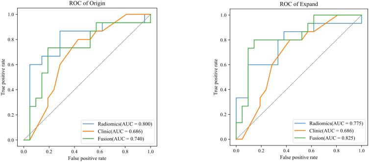
Results: The differences in HBeAg, capsule, target sign, and lymph node metastasis between MVI negative and positive groups were statistically significant (p < 0.05). Based on peritumoral, 1cm fusion model (in arterial phase) has an AUC of 0.940 (95% CI: 0.801-0.947) and 0.825 (95% CI: 0.633-0.917) in the training/testing set when identifying the MVI status of cHCC-CCA. The accuracy, sensitivity, and specificity in the testing set are 0.778, 0.800, and 0.726, respectively. The DCA shows that when the threshold is approximately 11.08%-66.47%, the net return of the fusion model is higher than that of the clinical and radiomics models under the same conditions.
Conclusion: Radiomics with a 1cm extension around the tumor can improve the performance of machine-learning models in predicting MVI labels.
 求助内容:
求助内容: 应助结果提醒方式:


