Josephine Warren, Daryl Cheng, Nigel Crawford, Bryn Jones, Rui Lun Ng, Annette Alafaci, Dion Stub, Philip Lew, Andrew J Taylor
{"title":"通过心脏磁共振成像提高COVID-19疫苗相关心肌炎合并心脏瘢痕的诊断","authors":"Josephine Warren, Daryl Cheng, Nigel Crawford, Bryn Jones, Rui Lun Ng, Annette Alafaci, Dion Stub, Philip Lew, Andrew J Taylor","doi":"10.1136/openhrt-2025-003333","DOIUrl":null,"url":null,"abstract":"<p><strong>Background: </strong>Myocarditis is a rare but potentially serious complication of COVID-19 vaccination. Cardiac magnetic resonance (CMR) with late gadolinium enhancement (LGE) imaging can identify cardiac scar, which may improve diagnostic accuracy and prognostication. We sought to define the incidence of long-term LGE post COVID-19 vaccine-associated myocarditis (C-VAM) and to establish the additive role of CMR in the diagnostic workup of this condition.</p><p><strong>Methods: </strong>Patients with Brighton Collaboration Criteria Level 1 (definite) or Level 2 (probable) C-VAM were prospectively recruited from the Surveillance of Adverse Events Following Vaccination In the Community database to undergo CMR at least 6 months after diagnosis. As there were limited patients with access to baseline CMR, prior CMR results were not included in the initial case definition. The presence of LGE at follow-up CMR was then integrated into the diagnostic algorithm, and the reclassification rate (definite vs probable) was calculated.</p><p><strong>Results: </strong>67 patients with C-VAM (mean age 30±13 years, 72% male) underwent CMR evaluation. The median time from vaccination to CMR was 548 (range 398-603) days. 20 patients (30%) had LGE. At diagnosis, nine patients (13%) were classified as definite and 58 (87%) as probable myocarditis. With the integration of CMR-LGE data, 16 patients (28%) were reclassified from probable to definite myocarditis.</p><p><strong>Conclusion: </strong>LGE on CMR occurred in one-third of patients with C-VAM. Without CMR at the time of diagnosis, almost one-third of patients are misclassified as probable rather than definite myocarditis, indicating a diagnostic strategy using echocardiography alone is insufficient.</p>","PeriodicalId":19505,"journal":{"name":"Open Heart","volume":"12 2","pages":""},"PeriodicalIF":2.8000,"publicationDate":"2025-07-18","publicationTypes":"Journal Article","fieldsOfStudy":null,"isOpenAccess":false,"openAccessPdf":"https://www.ncbi.nlm.nih.gov/pmc/articles/PMC12278148/pdf/","citationCount":"0","resultStr":"{\"title\":\"Improved diagnosis of COVID-19 vaccine-associated myocarditis with cardiac scarring identified by cardiac magnetic resonance imaging.\",\"authors\":\"Josephine Warren, Daryl Cheng, Nigel Crawford, Bryn Jones, Rui Lun Ng, Annette Alafaci, Dion Stub, Philip Lew, Andrew J Taylor\",\"doi\":\"10.1136/openhrt-2025-003333\",\"DOIUrl\":null,\"url\":null,\"abstract\":\"<p><strong>Background: </strong>Myocarditis is a rare but potentially serious complication of COVID-19 vaccination. Cardiac magnetic resonance (CMR) with late gadolinium enhancement (LGE) imaging can identify cardiac scar, which may improve diagnostic accuracy and prognostication. We sought to define the incidence of long-term LGE post COVID-19 vaccine-associated myocarditis (C-VAM) and to establish the additive role of CMR in the diagnostic workup of this condition.</p><p><strong>Methods: </strong>Patients with Brighton Collaboration Criteria Level 1 (definite) or Level 2 (probable) C-VAM were prospectively recruited from the Surveillance of Adverse Events Following Vaccination In the Community database to undergo CMR at least 6 months after diagnosis. As there were limited patients with access to baseline CMR, prior CMR results were not included in the initial case definition. The presence of LGE at follow-up CMR was then integrated into the diagnostic algorithm, and the reclassification rate (definite vs probable) was calculated.</p><p><strong>Results: </strong>67 patients with C-VAM (mean age 30±13 years, 72% male) underwent CMR evaluation. The median time from vaccination to CMR was 548 (range 398-603) days. 20 patients (30%) had LGE. At diagnosis, nine patients (13%) were classified as definite and 58 (87%) as probable myocarditis. With the integration of CMR-LGE data, 16 patients (28%) were reclassified from probable to definite myocarditis.</p><p><strong>Conclusion: </strong>LGE on CMR occurred in one-third of patients with C-VAM. Without CMR at the time of diagnosis, almost one-third of patients are misclassified as probable rather than definite myocarditis, indicating a diagnostic strategy using echocardiography alone is insufficient.</p>\",\"PeriodicalId\":19505,\"journal\":{\"name\":\"Open Heart\",\"volume\":\"12 2\",\"pages\":\"\"},\"PeriodicalIF\":2.8000,\"publicationDate\":\"2025-07-18\",\"publicationTypes\":\"Journal Article\",\"fieldsOfStudy\":null,\"isOpenAccess\":false,\"openAccessPdf\":\"https://www.ncbi.nlm.nih.gov/pmc/articles/PMC12278148/pdf/\",\"citationCount\":\"0\",\"resultStr\":null,\"platform\":\"Semanticscholar\",\"paperid\":null,\"PeriodicalName\":\"Open Heart\",\"FirstCategoryId\":\"1085\",\"ListUrlMain\":\"https://doi.org/10.1136/openhrt-2025-003333\",\"RegionNum\":0,\"RegionCategory\":null,\"ArticlePicture\":[],\"TitleCN\":null,\"AbstractTextCN\":null,\"PMCID\":null,\"EPubDate\":\"\",\"PubModel\":\"\",\"JCR\":\"Q2\",\"JCRName\":\"CARDIAC & CARDIOVASCULAR SYSTEMS\",\"Score\":null,\"Total\":0}","platform":"Semanticscholar","paperid":null,"PeriodicalName":"Open Heart","FirstCategoryId":"1085","ListUrlMain":"https://doi.org/10.1136/openhrt-2025-003333","RegionNum":0,"RegionCategory":null,"ArticlePicture":[],"TitleCN":null,"AbstractTextCN":null,"PMCID":null,"EPubDate":"","PubModel":"","JCR":"Q2","JCRName":"CARDIAC & CARDIOVASCULAR SYSTEMS","Score":null,"Total":0}
Improved diagnosis of COVID-19 vaccine-associated myocarditis with cardiac scarring identified by cardiac magnetic resonance imaging.
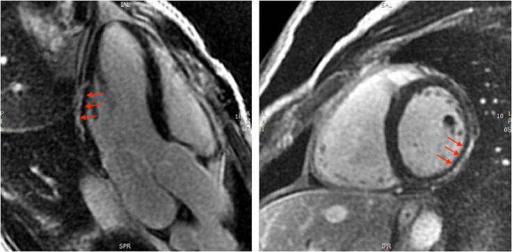
Background: Myocarditis is a rare but potentially serious complication of COVID-19 vaccination. Cardiac magnetic resonance (CMR) with late gadolinium enhancement (LGE) imaging can identify cardiac scar, which may improve diagnostic accuracy and prognostication. We sought to define the incidence of long-term LGE post COVID-19 vaccine-associated myocarditis (C-VAM) and to establish the additive role of CMR in the diagnostic workup of this condition.
Methods: Patients with Brighton Collaboration Criteria Level 1 (definite) or Level 2 (probable) C-VAM were prospectively recruited from the Surveillance of Adverse Events Following Vaccination In the Community database to undergo CMR at least 6 months after diagnosis. As there were limited patients with access to baseline CMR, prior CMR results were not included in the initial case definition. The presence of LGE at follow-up CMR was then integrated into the diagnostic algorithm, and the reclassification rate (definite vs probable) was calculated.
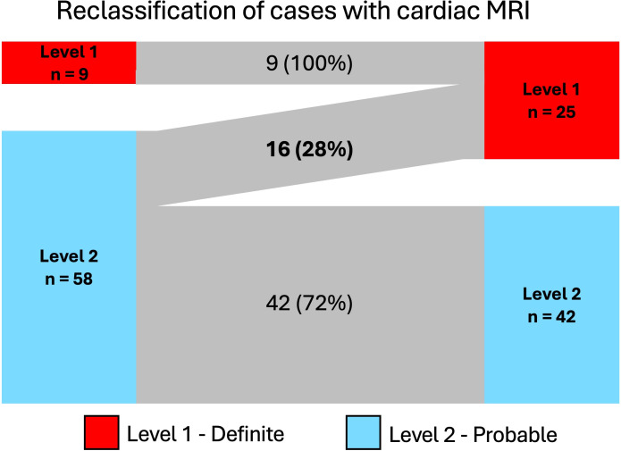
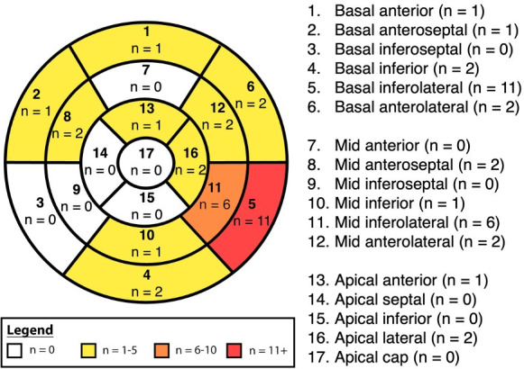
Results: 67 patients with C-VAM (mean age 30±13 years, 72% male) underwent CMR evaluation. The median time from vaccination to CMR was 548 (range 398-603) days. 20 patients (30%) had LGE. At diagnosis, nine patients (13%) were classified as definite and 58 (87%) as probable myocarditis. With the integration of CMR-LGE data, 16 patients (28%) were reclassified from probable to definite myocarditis.
Conclusion: LGE on CMR occurred in one-third of patients with C-VAM. Without CMR at the time of diagnosis, almost one-third of patients are misclassified as probable rather than definite myocarditis, indicating a diagnostic strategy using echocardiography alone is insufficient.
期刊介绍:
Open Heart is an online-only, open access cardiology journal that aims to be “open” in many ways: open access (free access for all readers), open peer review (unblinded peer review) and open data (data sharing is encouraged). The goal is to ensure maximum transparency and maximum impact on research progress and patient care. The journal is dedicated to publishing high quality, peer reviewed medical research in all disciplines and therapeutic areas of cardiovascular medicine. Research is published across all study phases and designs, from study protocols to phase I trials to meta-analyses, including small or specialist studies. Opinionated discussions on controversial topics are welcomed. Open Heart aims to operate a fast submission and review process with continuous publication online, to ensure timely, up-to-date research is available worldwide. The journal adheres to a rigorous and transparent peer review process, and all articles go through a statistical assessment to ensure robustness of the analyses. Open Heart is an official journal of the British Cardiovascular Society.

 求助内容:
求助内容: 应助结果提醒方式:


