Swagat Pradhan, Takayuki Mito, Nahid A Khan, Sofiia Olander, Aleksandra Zhaivoron, Thomas G McWilliams, Anu Suomalainen
{"title":"PDK4和营养反应解释了线粒体疾病中的肌肉特异性表现","authors":"Swagat Pradhan, Takayuki Mito, Nahid A Khan, Sofiia Olander, Aleksandra Zhaivoron, Thomas G McWilliams, Anu Suomalainen","doi":"10.1002/ctm2.70404","DOIUrl":null,"url":null,"abstract":"<div>\n \n \n <section>\n \n <h3> Background</h3>\n \n <p>Mitochondria elicit various metabolic stress responses, the roles of which in diseases are poorly understood. Here, we explore how different muscles of one individual—extraocular muscles (EOMs) and quadriceps femoris (QFs) muscles—respond to mitochondrial disease. The aim is to explain why EOMs atrophy early in the disease, unlike other muscles.</p>\n </section>\n \n <section>\n \n <h3> Methods</h3>\n \n <p>We used a mouse model for mitochondrial myopathy (“deletor”), which manifests progressive respiratory chain deficiency and human disease hallmarks in itsmuscles. Analyses included histology, ultrastructure, bulk and single-nuclear RNA-sequencing, metabolomics, and mitochondrial turnover assessed through in vivo mitophagy using transgenic mito-QC marker mice crossed to deletors.</p>\n </section>\n \n <section>\n \n <h3> Results</h3>\n \n <p>In mitochondrial muscle disease, large QFs upregulate glucose uptake that drives anabolic glycolytic one-carbon metabolism and mitochondrial integrated stress response. EOMs, however, react in an opposite manner, inhibiting glucose and pyruvate oxidation by activating PDK4, a pyruvate dehydrogenase kinase and inhibitor. Instead, EOMs upregulate acetyl-CoA synthesis and fatty-acid oxidation pathways, and accumulate lipids. In QFs, <i>Pdk4</i> transcription is not induced.- Amino acid levels are increased in QFs but are low in EOMs suggesting their catabolic use for energy metabolism. Mitophagy is stalled in both muscle types, in the most affected fibers.</p>\n </section>\n \n <section>\n \n <h3> Conclusions</h3>\n \n <p>Our evidence indicates that different muscles respond differently to mitochondrial disease even in one individual. While large muscles switch to anabolic mode and glycolysis, EOMs actively inhibit glucose usage. They upregulate lipid oxidation pathway, a non-optimal fuel choice in mitochondrial myopathy, leading to lipid accumulation and possibly increased reliance on amino acid oxidation. We propose that these consequences of non-optimal nutrient responses lead to EOMatrophy and progressive external ophthalmoplegia in patients. Our evidence highlights the importance of PDK4 and aberrant nutrient signaling underlying muscle atrophies.</p>\n </section>\n </div>","PeriodicalId":10189,"journal":{"name":"Clinical and Translational Medicine","volume":"15 7","pages":""},"PeriodicalIF":6.8000,"publicationDate":"2025-07-18","publicationTypes":"Journal Article","fieldsOfStudy":null,"isOpenAccess":false,"openAccessPdf":"https://onlinelibrary.wiley.com/doi/epdf/10.1002/ctm2.70404","citationCount":"0","resultStr":"{\"title\":\"PDK4 and nutrient responses explain muscle specific manifestation in mitochondrial disease\",\"authors\":\"Swagat Pradhan, Takayuki Mito, Nahid A Khan, Sofiia Olander, Aleksandra Zhaivoron, Thomas G McWilliams, Anu Suomalainen\",\"doi\":\"10.1002/ctm2.70404\",\"DOIUrl\":null,\"url\":null,\"abstract\":\"<div>\\n \\n \\n <section>\\n \\n <h3> Background</h3>\\n \\n <p>Mitochondria elicit various metabolic stress responses, the roles of which in diseases are poorly understood. Here, we explore how different muscles of one individual—extraocular muscles (EOMs) and quadriceps femoris (QFs) muscles—respond to mitochondrial disease. The aim is to explain why EOMs atrophy early in the disease, unlike other muscles.</p>\\n </section>\\n \\n <section>\\n \\n <h3> Methods</h3>\\n \\n <p>We used a mouse model for mitochondrial myopathy (“deletor”), which manifests progressive respiratory chain deficiency and human disease hallmarks in itsmuscles. Analyses included histology, ultrastructure, bulk and single-nuclear RNA-sequencing, metabolomics, and mitochondrial turnover assessed through in vivo mitophagy using transgenic mito-QC marker mice crossed to deletors.</p>\\n </section>\\n \\n <section>\\n \\n <h3> Results</h3>\\n \\n <p>In mitochondrial muscle disease, large QFs upregulate glucose uptake that drives anabolic glycolytic one-carbon metabolism and mitochondrial integrated stress response. EOMs, however, react in an opposite manner, inhibiting glucose and pyruvate oxidation by activating PDK4, a pyruvate dehydrogenase kinase and inhibitor. Instead, EOMs upregulate acetyl-CoA synthesis and fatty-acid oxidation pathways, and accumulate lipids. In QFs, <i>Pdk4</i> transcription is not induced.- Amino acid levels are increased in QFs but are low in EOMs suggesting their catabolic use for energy metabolism. Mitophagy is stalled in both muscle types, in the most affected fibers.</p>\\n </section>\\n \\n <section>\\n \\n <h3> Conclusions</h3>\\n \\n <p>Our evidence indicates that different muscles respond differently to mitochondrial disease even in one individual. While large muscles switch to anabolic mode and glycolysis, EOMs actively inhibit glucose usage. They upregulate lipid oxidation pathway, a non-optimal fuel choice in mitochondrial myopathy, leading to lipid accumulation and possibly increased reliance on amino acid oxidation. We propose that these consequences of non-optimal nutrient responses lead to EOMatrophy and progressive external ophthalmoplegia in patients. Our evidence highlights the importance of PDK4 and aberrant nutrient signaling underlying muscle atrophies.</p>\\n </section>\\n </div>\",\"PeriodicalId\":10189,\"journal\":{\"name\":\"Clinical and Translational Medicine\",\"volume\":\"15 7\",\"pages\":\"\"},\"PeriodicalIF\":6.8000,\"publicationDate\":\"2025-07-18\",\"publicationTypes\":\"Journal Article\",\"fieldsOfStudy\":null,\"isOpenAccess\":false,\"openAccessPdf\":\"https://onlinelibrary.wiley.com/doi/epdf/10.1002/ctm2.70404\",\"citationCount\":\"0\",\"resultStr\":null,\"platform\":\"Semanticscholar\",\"paperid\":null,\"PeriodicalName\":\"Clinical and Translational Medicine\",\"FirstCategoryId\":\"3\",\"ListUrlMain\":\"https://onlinelibrary.wiley.com/doi/10.1002/ctm2.70404\",\"RegionNum\":1,\"RegionCategory\":\"医学\",\"ArticlePicture\":[],\"TitleCN\":null,\"AbstractTextCN\":null,\"PMCID\":null,\"EPubDate\":\"\",\"PubModel\":\"\",\"JCR\":\"Q1\",\"JCRName\":\"MEDICINE, RESEARCH & EXPERIMENTAL\",\"Score\":null,\"Total\":0}","platform":"Semanticscholar","paperid":null,"PeriodicalName":"Clinical and Translational Medicine","FirstCategoryId":"3","ListUrlMain":"https://onlinelibrary.wiley.com/doi/10.1002/ctm2.70404","RegionNum":1,"RegionCategory":"医学","ArticlePicture":[],"TitleCN":null,"AbstractTextCN":null,"PMCID":null,"EPubDate":"","PubModel":"","JCR":"Q1","JCRName":"MEDICINE, RESEARCH & EXPERIMENTAL","Score":null,"Total":0}
PDK4 and nutrient responses explain muscle specific manifestation in mitochondrial disease
Background
Mitochondria elicit various metabolic stress responses, the roles of which in diseases are poorly understood. Here, we explore how different muscles of one individual—extraocular muscles (EOMs) and quadriceps femoris (QFs) muscles—respond to mitochondrial disease. The aim is to explain why EOMs atrophy early in the disease, unlike other muscles.
Methods
We used a mouse model for mitochondrial myopathy (“deletor”), which manifests progressive respiratory chain deficiency and human disease hallmarks in itsmuscles. Analyses included histology, ultrastructure, bulk and single-nuclear RNA-sequencing, metabolomics, and mitochondrial turnover assessed through in vivo mitophagy using transgenic mito-QC marker mice crossed to deletors.
Results
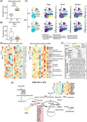
In mitochondrial muscle disease, large QFs upregulate glucose uptake that drives anabolic glycolytic one-carbon metabolism and mitochondrial integrated stress response. EOMs, however, react in an opposite manner, inhibiting glucose and pyruvate oxidation by activating PDK4, a pyruvate dehydrogenase kinase and inhibitor. Instead, EOMs upregulate acetyl-CoA synthesis and fatty-acid oxidation pathways, and accumulate lipids. In QFs, Pdk4 transcription is not induced.- Amino acid levels are increased in QFs but are low in EOMs suggesting their catabolic use for energy metabolism. Mitophagy is stalled in both muscle types, in the most affected fibers.
Conclusions
Our evidence indicates that different muscles respond differently to mitochondrial disease even in one individual. While large muscles switch to anabolic mode and glycolysis, EOMs actively inhibit glucose usage. They upregulate lipid oxidation pathway, a non-optimal fuel choice in mitochondrial myopathy, leading to lipid accumulation and possibly increased reliance on amino acid oxidation. We propose that these consequences of non-optimal nutrient responses lead to EOMatrophy and progressive external ophthalmoplegia in patients. Our evidence highlights the importance of PDK4 and aberrant nutrient signaling underlying muscle atrophies.
期刊介绍:
Clinical and Translational Medicine (CTM) is an international, peer-reviewed, open-access journal dedicated to accelerating the translation of preclinical research into clinical applications and fostering communication between basic and clinical scientists. It highlights the clinical potential and application of various fields including biotechnologies, biomaterials, bioengineering, biomarkers, molecular medicine, omics science, bioinformatics, immunology, molecular imaging, drug discovery, regulation, and health policy. With a focus on the bench-to-bedside approach, CTM prioritizes studies and clinical observations that generate hypotheses relevant to patients and diseases, guiding investigations in cellular and molecular medicine. The journal encourages submissions from clinicians, researchers, policymakers, and industry professionals.

 求助内容:
求助内容: 应助结果提醒方式:


