利用机器学习生物图像分析工具对睾丸组织学图像进行分割和量化;Ilastik和斐济软件
IF 1.9
Q2 MULTIDISCIPLINARY SCIENCES
引用次数: 0
摘要
组织形态学和组织化学技术广泛应用于不孕症研究,以评估睾丸损伤,确定相关机制,研究潜在的干预策略,监测治疗反应和预后。睾丸是男性的主要生殖器官,是由几个精小管和支持组织组成的区隔器官。因此,局灶性损伤是常见的,因此,要做出准确而深刻的推断,需要对几乎整个睾丸部分进行仔细的分析。然而,手工分析睾丸组织学切片以提取可量化的数据是忙碌的,耗时的,容易产生偏差和未检测到的斑块损伤和个人之间的可变性。为了规避这些挑战,我们使用免费的、开源的交互式基于机器学习的生物图像分析工具,提出了一个循序渐进的工作流程;伊拉斯蒂克和斐济。Ilastik使用随机森林分类器来计算用于图像分割的一般像素或对象特征。从ilastik输出的分割图像随后在斐济进行量化,以提取数据进行统计分析。•使用免费的,开源的交互式机器学习为基础的生物图像分析工具一步一步的工作流程;伊拉斯蒂克和斐济。•半自动化,可重复,节省时间,无偏,和广泛的方法分析和异质组织图像。•从图像中提取可量化的数据进行统计分析,得出综合结论。本文章由计算机程序翻译,如有差异,请以英文原文为准。
Segmentation and quantification of testicular histology images using machine learning bioimage analysis tools; Ilastik and Fiji software
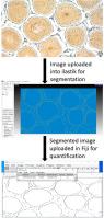
Histomorphological and histochemical techniques are widely used in infertility studies to assess testicular damage, determine the mechanisms involved, investigate potential interventions strategies, monitor treatment response and prognosis. Testis, a primary male reproductive organ is a compartmentalized organ made up of several seminiferous tubules and supporting tissue. Hence, focal damage is common, and accordingly, making accurate and insightful deductions require careful analysis of almost the entire testis section. However, manual analysis of testis histology sections to extract quantifiable data is hectic, time-consuming, liable to bias and undetected patchy damages and inter-personal variability. To circumvent these challenges, we present a step-by-step workflow using free, open-source interactive machine learning-based bioimage analysis tools; ilastik and Fiji. Ilastik uses a random forest classifier to compute generic pixel or object features for image segmentation. The segmented images exported from ilastik are subsequently quantified in FIJI to extract data for statistical analysis.
- •A step-by-step workflow using free, open-source interactive machine learning-based bioimage analysis tools; ilastik and Fiji.
- •A semiautomated, reproducible, time saving, unbiased, and broad scope method for analysis and heterogeneous tissue images.
- •Extraction of quantifiable data from images for statistical analysis to make comprehensive conclusions.
求助全文
通过发布文献求助,成功后即可免费获取论文全文。
去求助
来源期刊
MethodsX
Health Professions-Medical Laboratory Technology
CiteScore
3.60
自引率
5.30%
发文量
314
审稿时长
7 weeks
期刊介绍:
 求助内容:
求助内容: 应助结果提醒方式:


