{"title":"高尿酸血症与尾状核头部和壳核体积变小有关。","authors":"Naoki Omori, Manabu Ishida, Masahiro Takamura, Satoshi Abe, Atsushi Nagai","doi":"10.1093/braincomms/fcaf263","DOIUrl":null,"url":null,"abstract":"<p><p>Hyperuricaemia is a risk factor for gout, kidney diseases, and cerebrovascular diseases. Uric acid (UA) is also known as an antioxidant and has been suggested to inhibit the progression of neurodegenerative diseases, such as Parkinson's disease. However, only a few studies have focused on the potential effects of UA on the basal ganglia. This study aimed to measure UA levels and basal ganglia volumes in community-dwelling adults and to evaluate the association thereof. Blood UA levels and brain MRI data were collected from individuals who underwent brain health checkups between January 2015 and March 2022 at Shimane Institute of Health Science. Participants were classified into three groups based on their UA levels: normal-low UA (< 5.0 mg/dL), normal-high UA (5.0-7.0 mg/dL), and hyperuricaemia (> 7.0 mg/dL). MRI was used to assess the presence of asymptomatic infarcts, microbleeds, and the severity of enlarged perivascular spaces in the basal ganglia. The volumes of the caudate nucleus, globus pallidus, putamen, substantia nigra, and subthalamic nucleus were calculated using voxel-based morphometry (VBM) as <i>Z</i>-scores adjusted for participant age, sex, and total intracranial volume. Analysis of covariance and smooth-curve fitting models were used to examine the association between UA levels and basal ganglia volumes. In total, 981 participants were included in the analysis. Analysis of covariance revealed that the hyperuricaemia group had significantly higher <i>Z</i>-scores (indicating smaller volumes) in the bilateral caudate nucleus head and putamen than those of the normal-high UA group. The smooth-curve model showed U-shaped associations with smaller volumes for both high and low UA levels, whereas piecewise linear regression analysis confirmed significant regression lines only in the group with higher UA levels. Although UA is thought to have a neuroprotective effect in adults, our findings indicate that hyperuricaemia may contribute to smaller volumes in the caudate nucleus head and putamen. This suggests that excessive UA levels could negatively affect basal ganglia structure and neurological health.</p>","PeriodicalId":93915,"journal":{"name":"Brain communications","volume":"7 4","pages":"fcaf263"},"PeriodicalIF":4.5000,"publicationDate":"2025-07-15","publicationTypes":"Journal Article","fieldsOfStudy":null,"isOpenAccess":false,"openAccessPdf":"https://www.ncbi.nlm.nih.gov/pmc/articles/PMC12260223/pdf/","citationCount":"0","resultStr":"{\"title\":\"Hyperuricaemia is associated with smaller volumes in the caudate nucleus head and putamen.\",\"authors\":\"Naoki Omori, Manabu Ishida, Masahiro Takamura, Satoshi Abe, Atsushi Nagai\",\"doi\":\"10.1093/braincomms/fcaf263\",\"DOIUrl\":null,\"url\":null,\"abstract\":\"<p><p>Hyperuricaemia is a risk factor for gout, kidney diseases, and cerebrovascular diseases. Uric acid (UA) is also known as an antioxidant and has been suggested to inhibit the progression of neurodegenerative diseases, such as Parkinson's disease. However, only a few studies have focused on the potential effects of UA on the basal ganglia. This study aimed to measure UA levels and basal ganglia volumes in community-dwelling adults and to evaluate the association thereof. Blood UA levels and brain MRI data were collected from individuals who underwent brain health checkups between January 2015 and March 2022 at Shimane Institute of Health Science. Participants were classified into three groups based on their UA levels: normal-low UA (< 5.0 mg/dL), normal-high UA (5.0-7.0 mg/dL), and hyperuricaemia (> 7.0 mg/dL). MRI was used to assess the presence of asymptomatic infarcts, microbleeds, and the severity of enlarged perivascular spaces in the basal ganglia. The volumes of the caudate nucleus, globus pallidus, putamen, substantia nigra, and subthalamic nucleus were calculated using voxel-based morphometry (VBM) as <i>Z</i>-scores adjusted for participant age, sex, and total intracranial volume. Analysis of covariance and smooth-curve fitting models were used to examine the association between UA levels and basal ganglia volumes. In total, 981 participants were included in the analysis. Analysis of covariance revealed that the hyperuricaemia group had significantly higher <i>Z</i>-scores (indicating smaller volumes) in the bilateral caudate nucleus head and putamen than those of the normal-high UA group. The smooth-curve model showed U-shaped associations with smaller volumes for both high and low UA levels, whereas piecewise linear regression analysis confirmed significant regression lines only in the group with higher UA levels. Although UA is thought to have a neuroprotective effect in adults, our findings indicate that hyperuricaemia may contribute to smaller volumes in the caudate nucleus head and putamen. This suggests that excessive UA levels could negatively affect basal ganglia structure and neurological health.</p>\",\"PeriodicalId\":93915,\"journal\":{\"name\":\"Brain communications\",\"volume\":\"7 4\",\"pages\":\"fcaf263\"},\"PeriodicalIF\":4.5000,\"publicationDate\":\"2025-07-15\",\"publicationTypes\":\"Journal Article\",\"fieldsOfStudy\":null,\"isOpenAccess\":false,\"openAccessPdf\":\"https://www.ncbi.nlm.nih.gov/pmc/articles/PMC12260223/pdf/\",\"citationCount\":\"0\",\"resultStr\":null,\"platform\":\"Semanticscholar\",\"paperid\":null,\"PeriodicalName\":\"Brain communications\",\"FirstCategoryId\":\"1085\",\"ListUrlMain\":\"https://doi.org/10.1093/braincomms/fcaf263\",\"RegionNum\":0,\"RegionCategory\":null,\"ArticlePicture\":[],\"TitleCN\":null,\"AbstractTextCN\":null,\"PMCID\":null,\"EPubDate\":\"2025/1/1 0:00:00\",\"PubModel\":\"eCollection\",\"JCR\":\"Q1\",\"JCRName\":\"CLINICAL NEUROLOGY\",\"Score\":null,\"Total\":0}","platform":"Semanticscholar","paperid":null,"PeriodicalName":"Brain communications","FirstCategoryId":"1085","ListUrlMain":"https://doi.org/10.1093/braincomms/fcaf263","RegionNum":0,"RegionCategory":null,"ArticlePicture":[],"TitleCN":null,"AbstractTextCN":null,"PMCID":null,"EPubDate":"2025/1/1 0:00:00","PubModel":"eCollection","JCR":"Q1","JCRName":"CLINICAL NEUROLOGY","Score":null,"Total":0}
Hyperuricaemia is associated with smaller volumes in the caudate nucleus head and putamen.
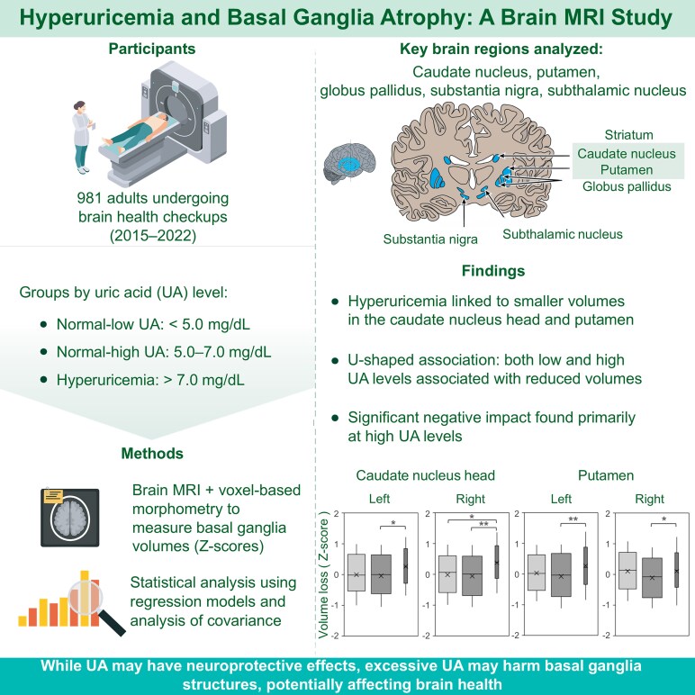
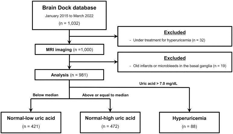
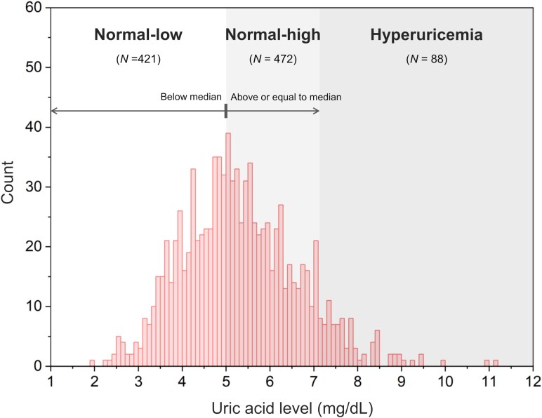
Hyperuricaemia is a risk factor for gout, kidney diseases, and cerebrovascular diseases. Uric acid (UA) is also known as an antioxidant and has been suggested to inhibit the progression of neurodegenerative diseases, such as Parkinson's disease. However, only a few studies have focused on the potential effects of UA on the basal ganglia. This study aimed to measure UA levels and basal ganglia volumes in community-dwelling adults and to evaluate the association thereof. Blood UA levels and brain MRI data were collected from individuals who underwent brain health checkups between January 2015 and March 2022 at Shimane Institute of Health Science. Participants were classified into three groups based on their UA levels: normal-low UA (< 5.0 mg/dL), normal-high UA (5.0-7.0 mg/dL), and hyperuricaemia (> 7.0 mg/dL). MRI was used to assess the presence of asymptomatic infarcts, microbleeds, and the severity of enlarged perivascular spaces in the basal ganglia. The volumes of the caudate nucleus, globus pallidus, putamen, substantia nigra, and subthalamic nucleus were calculated using voxel-based morphometry (VBM) as Z-scores adjusted for participant age, sex, and total intracranial volume. Analysis of covariance and smooth-curve fitting models were used to examine the association between UA levels and basal ganglia volumes. In total, 981 participants were included in the analysis. Analysis of covariance revealed that the hyperuricaemia group had significantly higher Z-scores (indicating smaller volumes) in the bilateral caudate nucleus head and putamen than those of the normal-high UA group. The smooth-curve model showed U-shaped associations with smaller volumes for both high and low UA levels, whereas piecewise linear regression analysis confirmed significant regression lines only in the group with higher UA levels. Although UA is thought to have a neuroprotective effect in adults, our findings indicate that hyperuricaemia may contribute to smaller volumes in the caudate nucleus head and putamen. This suggests that excessive UA levels could negatively affect basal ganglia structure and neurological health.

 求助内容:
求助内容: 应助结果提醒方式:


