Patricia M Sikorski, Henry J Kaminski, Linda L Kusner
{"title":"重症肌无力患者胸腺增生:述评。","authors":"Patricia M Sikorski, Henry J Kaminski, Linda L Kusner","doi":"10.21037/med-25-12","DOIUrl":null,"url":null,"abstract":"<p><strong>Background and objective: </strong>Thymic pathology is observed in approximately 80% of patients with acetylcholine receptor antibody-positive myasthenia gravis (AChR-MG). Among these thymic abnormalities, thymic follicular hyperplasia (TFH) is commonly associated with early-onset MG (EOMG). TFH is characterized by the presence of lymphoid follicles and germinal center (GC) formation, which are closely linked with the breakdown of tolerance to the AChR. GCs promote the development of autoreactive plasma cells that secrete autoantibodies against the AChR, contributing to the disease pathology. In this review, we examine current knowledge on thymic pathology in EOMG, immunological and environmental factors contributing to the development of thymic hyperplasia and highlight avenues for future research.</p><p><strong>Methods: </strong>A comprehensive literature search was conducted using PubMed without restriction on publication date. Articles were included if they discussed the function of the thymus, thymic pathology in MG, thymic hyperplasia in EOMG, or focused on cellular or molecular mechanisms associated with TFH in EOMG.</p><p><strong>Key content and findings: </strong>TFH is a hallmark of EOMG, characterized by GC formation and intrathymic production of AChR autoantibodies. The hyperplastic thymus exhibits heightened interferon (IFN) signaling, toll-like receptor (TLR) activation, altered chemokine expression, accumulation of B and T cells, and expression of AChR by thymic epithelial cells, creating a pro-autoimmune environment. Sex-related differences, particularly estrogen's effects on autoimmune regulator gene (AIRE) expression and immune tolerance, may contribute to the female predominance in EOMG. Emerging technologies such as single-cell and spatial transcriptomics, along with thymic organoid models, offer new avenues to study the mechanisms driving TFH.</p><p><strong>Conclusions: </strong>TFH reflects the convergence of immune dysregulation and structural abnormalities that together promote the loss of tolerance in EOMG. The interindividual variability in thymic pathology and treatment response underscores the need for more personalized therapeutic strategies. Advances in high-resolution profiling and experimental modeling will be essential to uncover the underlying drivers of thymic hyperplasia and to guide the development of targeted therapies for MG.</p>","PeriodicalId":74139,"journal":{"name":"Mediastinum (Hong Kong, China)","volume":"9 ","pages":"17"},"PeriodicalIF":0.0000,"publicationDate":"2025-06-25","publicationTypes":"Journal Article","fieldsOfStudy":null,"isOpenAccess":false,"openAccessPdf":"https://www.ncbi.nlm.nih.gov/pmc/articles/PMC12260958/pdf/","citationCount":"0","resultStr":"{\"title\":\"Thymic hyperplasia in myasthenia gravis: a narrative review.\",\"authors\":\"Patricia M Sikorski, Henry J Kaminski, Linda L Kusner\",\"doi\":\"10.21037/med-25-12\",\"DOIUrl\":null,\"url\":null,\"abstract\":\"<p><strong>Background and objective: </strong>Thymic pathology is observed in approximately 80% of patients with acetylcholine receptor antibody-positive myasthenia gravis (AChR-MG). Among these thymic abnormalities, thymic follicular hyperplasia (TFH) is commonly associated with early-onset MG (EOMG). TFH is characterized by the presence of lymphoid follicles and germinal center (GC) formation, which are closely linked with the breakdown of tolerance to the AChR. GCs promote the development of autoreactive plasma cells that secrete autoantibodies against the AChR, contributing to the disease pathology. In this review, we examine current knowledge on thymic pathology in EOMG, immunological and environmental factors contributing to the development of thymic hyperplasia and highlight avenues for future research.</p><p><strong>Methods: </strong>A comprehensive literature search was conducted using PubMed without restriction on publication date. Articles were included if they discussed the function of the thymus, thymic pathology in MG, thymic hyperplasia in EOMG, or focused on cellular or molecular mechanisms associated with TFH in EOMG.</p><p><strong>Key content and findings: </strong>TFH is a hallmark of EOMG, characterized by GC formation and intrathymic production of AChR autoantibodies. The hyperplastic thymus exhibits heightened interferon (IFN) signaling, toll-like receptor (TLR) activation, altered chemokine expression, accumulation of B and T cells, and expression of AChR by thymic epithelial cells, creating a pro-autoimmune environment. Sex-related differences, particularly estrogen's effects on autoimmune regulator gene (AIRE) expression and immune tolerance, may contribute to the female predominance in EOMG. Emerging technologies such as single-cell and spatial transcriptomics, along with thymic organoid models, offer new avenues to study the mechanisms driving TFH.</p><p><strong>Conclusions: </strong>TFH reflects the convergence of immune dysregulation and structural abnormalities that together promote the loss of tolerance in EOMG. The interindividual variability in thymic pathology and treatment response underscores the need for more personalized therapeutic strategies. Advances in high-resolution profiling and experimental modeling will be essential to uncover the underlying drivers of thymic hyperplasia and to guide the development of targeted therapies for MG.</p>\",\"PeriodicalId\":74139,\"journal\":{\"name\":\"Mediastinum (Hong Kong, China)\",\"volume\":\"9 \",\"pages\":\"17\"},\"PeriodicalIF\":0.0000,\"publicationDate\":\"2025-06-25\",\"publicationTypes\":\"Journal Article\",\"fieldsOfStudy\":null,\"isOpenAccess\":false,\"openAccessPdf\":\"https://www.ncbi.nlm.nih.gov/pmc/articles/PMC12260958/pdf/\",\"citationCount\":\"0\",\"resultStr\":null,\"platform\":\"Semanticscholar\",\"paperid\":null,\"PeriodicalName\":\"Mediastinum (Hong Kong, China)\",\"FirstCategoryId\":\"1085\",\"ListUrlMain\":\"https://doi.org/10.21037/med-25-12\",\"RegionNum\":0,\"RegionCategory\":null,\"ArticlePicture\":[],\"TitleCN\":null,\"AbstractTextCN\":null,\"PMCID\":null,\"EPubDate\":\"2025/1/1 0:00:00\",\"PubModel\":\"eCollection\",\"JCR\":\"\",\"JCRName\":\"\",\"Score\":null,\"Total\":0}","platform":"Semanticscholar","paperid":null,"PeriodicalName":"Mediastinum (Hong Kong, China)","FirstCategoryId":"1085","ListUrlMain":"https://doi.org/10.21037/med-25-12","RegionNum":0,"RegionCategory":null,"ArticlePicture":[],"TitleCN":null,"AbstractTextCN":null,"PMCID":null,"EPubDate":"2025/1/1 0:00:00","PubModel":"eCollection","JCR":"","JCRName":"","Score":null,"Total":0}
Thymic hyperplasia in myasthenia gravis: a narrative review.
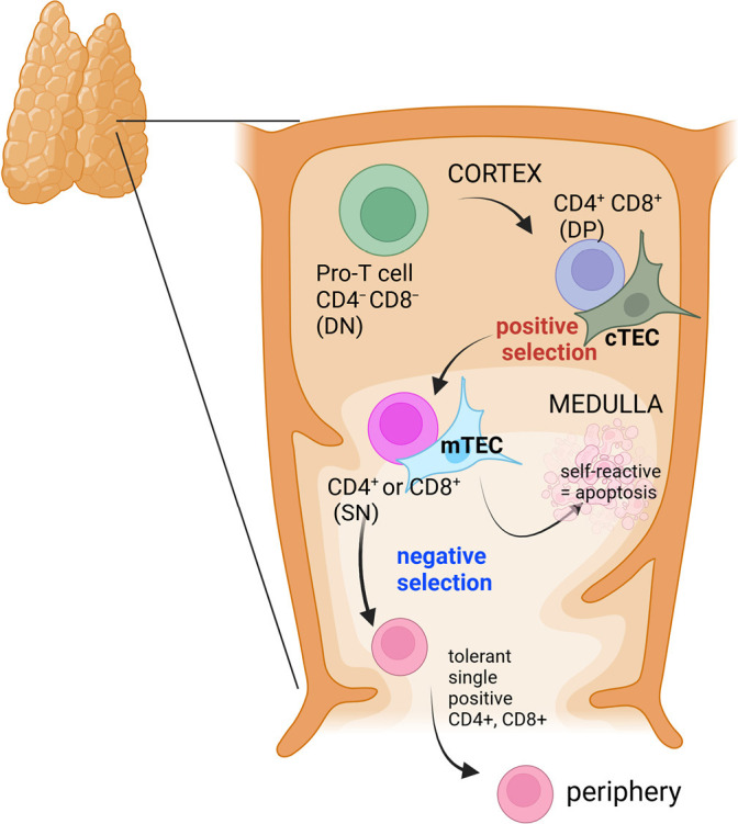
Background and objective: Thymic pathology is observed in approximately 80% of patients with acetylcholine receptor antibody-positive myasthenia gravis (AChR-MG). Among these thymic abnormalities, thymic follicular hyperplasia (TFH) is commonly associated with early-onset MG (EOMG). TFH is characterized by the presence of lymphoid follicles and germinal center (GC) formation, which are closely linked with the breakdown of tolerance to the AChR. GCs promote the development of autoreactive plasma cells that secrete autoantibodies against the AChR, contributing to the disease pathology. In this review, we examine current knowledge on thymic pathology in EOMG, immunological and environmental factors contributing to the development of thymic hyperplasia and highlight avenues for future research.
Methods: A comprehensive literature search was conducted using PubMed without restriction on publication date. Articles were included if they discussed the function of the thymus, thymic pathology in MG, thymic hyperplasia in EOMG, or focused on cellular or molecular mechanisms associated with TFH in EOMG.
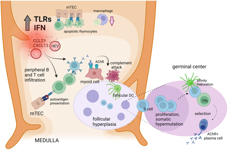
Key content and findings: TFH is a hallmark of EOMG, characterized by GC formation and intrathymic production of AChR autoantibodies. The hyperplastic thymus exhibits heightened interferon (IFN) signaling, toll-like receptor (TLR) activation, altered chemokine expression, accumulation of B and T cells, and expression of AChR by thymic epithelial cells, creating a pro-autoimmune environment. Sex-related differences, particularly estrogen's effects on autoimmune regulator gene (AIRE) expression and immune tolerance, may contribute to the female predominance in EOMG. Emerging technologies such as single-cell and spatial transcriptomics, along with thymic organoid models, offer new avenues to study the mechanisms driving TFH.
Conclusions: TFH reflects the convergence of immune dysregulation and structural abnormalities that together promote the loss of tolerance in EOMG. The interindividual variability in thymic pathology and treatment response underscores the need for more personalized therapeutic strategies. Advances in high-resolution profiling and experimental modeling will be essential to uncover the underlying drivers of thymic hyperplasia and to guide the development of targeted therapies for MG.

 求助内容:
求助内容: 应助结果提醒方式:


