Jackson S Hamersly, Mason D Tippy, James E Slaven, Yohan Jang, Lauren M Ladd, Mark T Dillon
{"title":"骨折后逆行全肩关节置换术患者的肩关节形态。","authors":"Jackson S Hamersly, Mason D Tippy, James E Slaven, Yohan Jang, Lauren M Ladd, Mark T Dillon","doi":"10.1007/s00402-025-05977-8","DOIUrl":null,"url":null,"abstract":"<p><strong>Introduction: </strong>Glenoid morphology in patients undergoing reverse total shoulder arthroplasty (rTSA) due to arthritis has been previously studied; however, it has not been as thoroughly evaluated in fracture populations. The purpose of this study is to utilize pre-operative computed tomography (CT) scans to better understand the glenoid anatomy of those patients undergoing rTSA due to fracture.</p><p><strong>Materials and methods: </strong>Patients over the age of 18 who underwent rTSA for proximal humerus fractures from January 1, 2015 to October 31, 2023 at two university health system affiliated hospitals were included if they had a CT scan available for review and image reconstruction. Patients were excluded if a pathologic fracture was identified, surgery was performed greater than 6 weeks after the initial injury, surgery was a conversion or revision surgery, or if a glenoid fracture was present. Glenoid version and reverse shoulder arthroplasty (RSA) angles were measured by a musculoskeletal fellowship-trained radiologist and a shoulder and elbow fellowship-trained orthopaedic surgeon and averaged for final values. Glenoid morphologies were determined using the Walch and Favard classifications.</p><p><strong>Results: </strong>A total of 53 patients with a mean age of 70.4 years (range 36.6-91.2) were included in this study, 84.9% of which were female. Walch A1 glenoid morphology was noted in 92.5% of patients, and Favard E0 morphology was present in 98.1% of patients. Median glenoid version was 3° of retroversion. Median RSA angle was 19°. Of note, 37.7% of patients had a RSA angle of ≥ 20°.</p><p><strong>Conclusions: </strong>Patients undergoing rTSA for fracture may not have significant glenoid deformity from arthritic wear. However, surgeons should be aware of variations in glenoid version and RSA angle. In this study population, over one-third of patients had a RSA angle of ≥ 20°. Thus, surgeons should take these findings into account when performing rTSA for fracture.</p>","PeriodicalId":8326,"journal":{"name":"Archives of Orthopaedic and Trauma Surgery","volume":"145 1","pages":"375"},"PeriodicalIF":2.1000,"publicationDate":"2025-07-15","publicationTypes":"Journal Article","fieldsOfStudy":null,"isOpenAccess":false,"openAccessPdf":"https://www.ncbi.nlm.nih.gov/pmc/articles/PMC12263477/pdf/","citationCount":"0","resultStr":"{\"title\":\"Glenoid morphology in patients undergoing reverse total shoulder arthroplasty due to fracture.\",\"authors\":\"Jackson S Hamersly, Mason D Tippy, James E Slaven, Yohan Jang, Lauren M Ladd, Mark T Dillon\",\"doi\":\"10.1007/s00402-025-05977-8\",\"DOIUrl\":null,\"url\":null,\"abstract\":\"<p><strong>Introduction: </strong>Glenoid morphology in patients undergoing reverse total shoulder arthroplasty (rTSA) due to arthritis has been previously studied; however, it has not been as thoroughly evaluated in fracture populations. The purpose of this study is to utilize pre-operative computed tomography (CT) scans to better understand the glenoid anatomy of those patients undergoing rTSA due to fracture.</p><p><strong>Materials and methods: </strong>Patients over the age of 18 who underwent rTSA for proximal humerus fractures from January 1, 2015 to October 31, 2023 at two university health system affiliated hospitals were included if they had a CT scan available for review and image reconstruction. Patients were excluded if a pathologic fracture was identified, surgery was performed greater than 6 weeks after the initial injury, surgery was a conversion or revision surgery, or if a glenoid fracture was present. Glenoid version and reverse shoulder arthroplasty (RSA) angles were measured by a musculoskeletal fellowship-trained radiologist and a shoulder and elbow fellowship-trained orthopaedic surgeon and averaged for final values. Glenoid morphologies were determined using the Walch and Favard classifications.</p><p><strong>Results: </strong>A total of 53 patients with a mean age of 70.4 years (range 36.6-91.2) were included in this study, 84.9% of which were female. Walch A1 glenoid morphology was noted in 92.5% of patients, and Favard E0 morphology was present in 98.1% of patients. Median glenoid version was 3° of retroversion. Median RSA angle was 19°. Of note, 37.7% of patients had a RSA angle of ≥ 20°.</p><p><strong>Conclusions: </strong>Patients undergoing rTSA for fracture may not have significant glenoid deformity from arthritic wear. However, surgeons should be aware of variations in glenoid version and RSA angle. In this study population, over one-third of patients had a RSA angle of ≥ 20°. Thus, surgeons should take these findings into account when performing rTSA for fracture.</p>\",\"PeriodicalId\":8326,\"journal\":{\"name\":\"Archives of Orthopaedic and Trauma Surgery\",\"volume\":\"145 1\",\"pages\":\"375\"},\"PeriodicalIF\":2.1000,\"publicationDate\":\"2025-07-15\",\"publicationTypes\":\"Journal Article\",\"fieldsOfStudy\":null,\"isOpenAccess\":false,\"openAccessPdf\":\"https://www.ncbi.nlm.nih.gov/pmc/articles/PMC12263477/pdf/\",\"citationCount\":\"0\",\"resultStr\":null,\"platform\":\"Semanticscholar\",\"paperid\":null,\"PeriodicalName\":\"Archives of Orthopaedic and Trauma Surgery\",\"FirstCategoryId\":\"3\",\"ListUrlMain\":\"https://doi.org/10.1007/s00402-025-05977-8\",\"RegionNum\":3,\"RegionCategory\":\"医学\",\"ArticlePicture\":[],\"TitleCN\":null,\"AbstractTextCN\":null,\"PMCID\":null,\"EPubDate\":\"\",\"PubModel\":\"\",\"JCR\":\"Q2\",\"JCRName\":\"ORTHOPEDICS\",\"Score\":null,\"Total\":0}","platform":"Semanticscholar","paperid":null,"PeriodicalName":"Archives of Orthopaedic and Trauma Surgery","FirstCategoryId":"3","ListUrlMain":"https://doi.org/10.1007/s00402-025-05977-8","RegionNum":3,"RegionCategory":"医学","ArticlePicture":[],"TitleCN":null,"AbstractTextCN":null,"PMCID":null,"EPubDate":"","PubModel":"","JCR":"Q2","JCRName":"ORTHOPEDICS","Score":null,"Total":0}
Glenoid morphology in patients undergoing reverse total shoulder arthroplasty due to fracture.
Introduction: Glenoid morphology in patients undergoing reverse total shoulder arthroplasty (rTSA) due to arthritis has been previously studied; however, it has not been as thoroughly evaluated in fracture populations. The purpose of this study is to utilize pre-operative computed tomography (CT) scans to better understand the glenoid anatomy of those patients undergoing rTSA due to fracture.
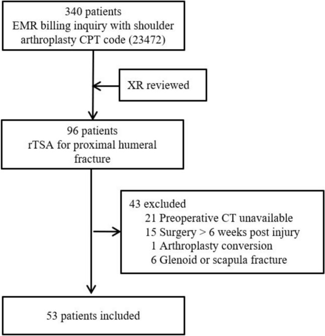
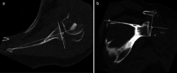
Materials and methods: Patients over the age of 18 who underwent rTSA for proximal humerus fractures from January 1, 2015 to October 31, 2023 at two university health system affiliated hospitals were included if they had a CT scan available for review and image reconstruction. Patients were excluded if a pathologic fracture was identified, surgery was performed greater than 6 weeks after the initial injury, surgery was a conversion or revision surgery, or if a glenoid fracture was present. Glenoid version and reverse shoulder arthroplasty (RSA) angles were measured by a musculoskeletal fellowship-trained radiologist and a shoulder and elbow fellowship-trained orthopaedic surgeon and averaged for final values. Glenoid morphologies were determined using the Walch and Favard classifications.
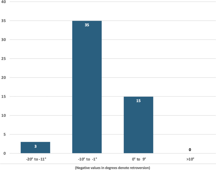
Results: A total of 53 patients with a mean age of 70.4 years (range 36.6-91.2) were included in this study, 84.9% of which were female. Walch A1 glenoid morphology was noted in 92.5% of patients, and Favard E0 morphology was present in 98.1% of patients. Median glenoid version was 3° of retroversion. Median RSA angle was 19°. Of note, 37.7% of patients had a RSA angle of ≥ 20°.
Conclusions: Patients undergoing rTSA for fracture may not have significant glenoid deformity from arthritic wear. However, surgeons should be aware of variations in glenoid version and RSA angle. In this study population, over one-third of patients had a RSA angle of ≥ 20°. Thus, surgeons should take these findings into account when performing rTSA for fracture.
期刊介绍:
"Archives of Orthopaedic and Trauma Surgery" is a rich source of instruction and information for physicians in clinical practice and research in the extensive field of orthopaedics and traumatology. The journal publishes papers that deal with diseases and injuries of the musculoskeletal system from all fields and aspects of medicine. The journal is particularly interested in papers that satisfy the information needs of orthopaedic clinicians and practitioners. The journal places special emphasis on clinical relevance.
"Archives of Orthopaedic and Trauma Surgery" is the official journal of the German Speaking Arthroscopy Association (AGA).
 求助内容:
求助内容: 应助结果提醒方式:


