{"title":"18f -前列腺特异性膜抗原-1007 PET/MRI与18f -前列腺特异性膜抗原-1007 PET/计算机断层扫描对前列腺癌根治性切除术后生化复发的诊断价值","authors":"Yuping Zeng, Hengbin Liao, Guihua Jiang, Gongfa Wu, Junyuan Zhong","doi":"10.1097/MNM.0000000000002028","DOIUrl":null,"url":null,"abstract":"<p><strong>Objective: </strong>To assess the diagnostic value of fluorine 18 ( 18 F)-labeled prostate-specific membrane antigen (PSMA)-1007 PET/MRI and compare with that of 18 F-PSMA-1007 PET/computed tomography (CT) for biochemical recurrence (BCR) of prostate cancer (PCa) after radical prostatectomy.</p><p><strong>Materials and methods: </strong>We enrolled 40 patients who underwent 18 F-PSMA-1007 PET/CT and 18 F-PSMA-1007 PET/MRI for BCR after radical prostatectomy. Two readers independently assessed the images and determined their overall assessment of positive lesions on PET/MRI and PET/CT. The association between the patients' clinical characteristics and positive detection results on the PET/CT and PET/MRI was explored. The PET/CT and PET/MRI results were verified during a 24-month follow-up to calculate their diagnostic accuracy.</p><p><strong>Results: </strong>The detection rate of positive patients on 18 F-PSMA-1007 PET/CT and PET/MRI were consistent, with a value of 77.50%. The positive detection results were moderately associated with the patients' prostate-specific antigen (PSA) levels at examination, and the detection rate increased significantly with values of 50.00%, 72.73%, 90.91%, and 100% for PSA levels <0.5, 0.5-<1.0, 1.0-<2.0, and ≥2.0 ng/ml, respectively. Conclusive follow-up for affirmation or refutation of PCa recurrence was available for 33 patients. Compared with the follow-up results, on the patient-based level, the diagnostic accuracies for PET/MRI and PET/CT were both 100%. On the lesion-based level, PET/MRI excluded a false positive of bone metastasis on PET/CT.</p><p><strong>Conclusion: </strong>18 F-PSMA-1007 PET/MRI and 18 F-PSMA-1007 PET/CT demonstrate almost equal diagnostic value in detecting BCR, but PET/MRI can provide more lesion information, facilitating diagnosis and treatment due to its superior soft tissue resolution.</p>","PeriodicalId":19708,"journal":{"name":"Nuclear Medicine Communications","volume":" ","pages":"1052-1060"},"PeriodicalIF":1.3000,"publicationDate":"2025-11-01","publicationTypes":"Journal Article","fieldsOfStudy":null,"isOpenAccess":false,"openAccessPdf":"https://www.ncbi.nlm.nih.gov/pmc/articles/PMC12502934/pdf/","citationCount":"0","resultStr":"{\"title\":\"Diagnostic value of 18 F-prostate-specific membrane antigen-1007 PET/MRI versus 18 F-prostate-specific membrane antigen-1007 PET/computed tomography for biochemical recurrence of prostate cancer after radical prostatectomy.\",\"authors\":\"Yuping Zeng, Hengbin Liao, Guihua Jiang, Gongfa Wu, Junyuan Zhong\",\"doi\":\"10.1097/MNM.0000000000002028\",\"DOIUrl\":null,\"url\":null,\"abstract\":\"<p><strong>Objective: </strong>To assess the diagnostic value of fluorine 18 ( 18 F)-labeled prostate-specific membrane antigen (PSMA)-1007 PET/MRI and compare with that of 18 F-PSMA-1007 PET/computed tomography (CT) for biochemical recurrence (BCR) of prostate cancer (PCa) after radical prostatectomy.</p><p><strong>Materials and methods: </strong>We enrolled 40 patients who underwent 18 F-PSMA-1007 PET/CT and 18 F-PSMA-1007 PET/MRI for BCR after radical prostatectomy. Two readers independently assessed the images and determined their overall assessment of positive lesions on PET/MRI and PET/CT. The association between the patients' clinical characteristics and positive detection results on the PET/CT and PET/MRI was explored. The PET/CT and PET/MRI results were verified during a 24-month follow-up to calculate their diagnostic accuracy.</p><p><strong>Results: </strong>The detection rate of positive patients on 18 F-PSMA-1007 PET/CT and PET/MRI were consistent, with a value of 77.50%. The positive detection results were moderately associated with the patients' prostate-specific antigen (PSA) levels at examination, and the detection rate increased significantly with values of 50.00%, 72.73%, 90.91%, and 100% for PSA levels <0.5, 0.5-<1.0, 1.0-<2.0, and ≥2.0 ng/ml, respectively. Conclusive follow-up for affirmation or refutation of PCa recurrence was available for 33 patients. Compared with the follow-up results, on the patient-based level, the diagnostic accuracies for PET/MRI and PET/CT were both 100%. On the lesion-based level, PET/MRI excluded a false positive of bone metastasis on PET/CT.</p><p><strong>Conclusion: </strong>18 F-PSMA-1007 PET/MRI and 18 F-PSMA-1007 PET/CT demonstrate almost equal diagnostic value in detecting BCR, but PET/MRI can provide more lesion information, facilitating diagnosis and treatment due to its superior soft tissue resolution.</p>\",\"PeriodicalId\":19708,\"journal\":{\"name\":\"Nuclear Medicine Communications\",\"volume\":\" \",\"pages\":\"1052-1060\"},\"PeriodicalIF\":1.3000,\"publicationDate\":\"2025-11-01\",\"publicationTypes\":\"Journal Article\",\"fieldsOfStudy\":null,\"isOpenAccess\":false,\"openAccessPdf\":\"https://www.ncbi.nlm.nih.gov/pmc/articles/PMC12502934/pdf/\",\"citationCount\":\"0\",\"resultStr\":null,\"platform\":\"Semanticscholar\",\"paperid\":null,\"PeriodicalName\":\"Nuclear Medicine Communications\",\"FirstCategoryId\":\"3\",\"ListUrlMain\":\"https://doi.org/10.1097/MNM.0000000000002028\",\"RegionNum\":4,\"RegionCategory\":\"医学\",\"ArticlePicture\":[],\"TitleCN\":null,\"AbstractTextCN\":null,\"PMCID\":null,\"EPubDate\":\"2025/7/15 0:00:00\",\"PubModel\":\"Epub\",\"JCR\":\"Q3\",\"JCRName\":\"RADIOLOGY, NUCLEAR MEDICINE & MEDICAL IMAGING\",\"Score\":null,\"Total\":0}","platform":"Semanticscholar","paperid":null,"PeriodicalName":"Nuclear Medicine Communications","FirstCategoryId":"3","ListUrlMain":"https://doi.org/10.1097/MNM.0000000000002028","RegionNum":4,"RegionCategory":"医学","ArticlePicture":[],"TitleCN":null,"AbstractTextCN":null,"PMCID":null,"EPubDate":"2025/7/15 0:00:00","PubModel":"Epub","JCR":"Q3","JCRName":"RADIOLOGY, NUCLEAR MEDICINE & MEDICAL IMAGING","Score":null,"Total":0}
Diagnostic value of 18 F-prostate-specific membrane antigen-1007 PET/MRI versus 18 F-prostate-specific membrane antigen-1007 PET/computed tomography for biochemical recurrence of prostate cancer after radical prostatectomy.
Objective: To assess the diagnostic value of fluorine 18 ( 18 F)-labeled prostate-specific membrane antigen (PSMA)-1007 PET/MRI and compare with that of 18 F-PSMA-1007 PET/computed tomography (CT) for biochemical recurrence (BCR) of prostate cancer (PCa) after radical prostatectomy.
Materials and methods: We enrolled 40 patients who underwent 18 F-PSMA-1007 PET/CT and 18 F-PSMA-1007 PET/MRI for BCR after radical prostatectomy. Two readers independently assessed the images and determined their overall assessment of positive lesions on PET/MRI and PET/CT. The association between the patients' clinical characteristics and positive detection results on the PET/CT and PET/MRI was explored. The PET/CT and PET/MRI results were verified during a 24-month follow-up to calculate their diagnostic accuracy.
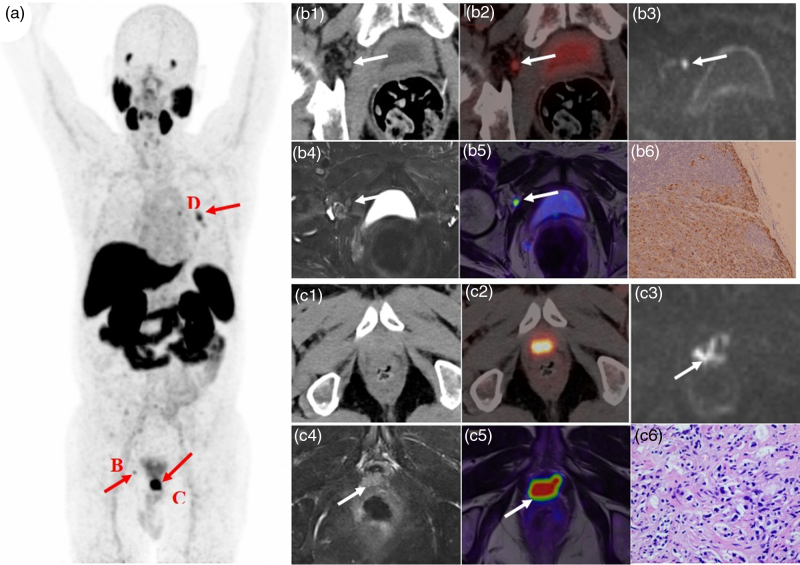
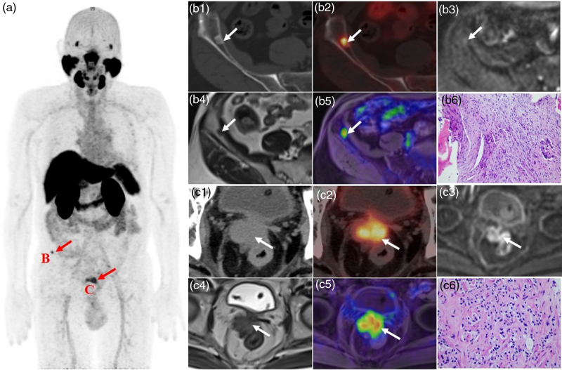
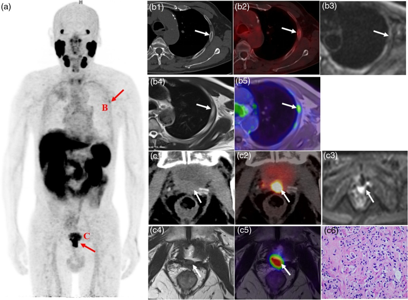
Results: The detection rate of positive patients on 18 F-PSMA-1007 PET/CT and PET/MRI were consistent, with a value of 77.50%. The positive detection results were moderately associated with the patients' prostate-specific antigen (PSA) levels at examination, and the detection rate increased significantly with values of 50.00%, 72.73%, 90.91%, and 100% for PSA levels <0.5, 0.5-<1.0, 1.0-<2.0, and ≥2.0 ng/ml, respectively. Conclusive follow-up for affirmation or refutation of PCa recurrence was available for 33 patients. Compared with the follow-up results, on the patient-based level, the diagnostic accuracies for PET/MRI and PET/CT were both 100%. On the lesion-based level, PET/MRI excluded a false positive of bone metastasis on PET/CT.
Conclusion: 18 F-PSMA-1007 PET/MRI and 18 F-PSMA-1007 PET/CT demonstrate almost equal diagnostic value in detecting BCR, but PET/MRI can provide more lesion information, facilitating diagnosis and treatment due to its superior soft tissue resolution.
期刊介绍:
Nuclear Medicine Communications, the official journal of the British Nuclear Medicine Society, is a rapid communications journal covering nuclear medicine and molecular imaging with radionuclides, and the basic supporting sciences. As well as clinical research and commentary, manuscripts describing research on preclinical and basic sciences (radiochemistry, radiopharmacy, radiobiology, radiopharmacology, medical physics, computing and engineering, and technical and nursing professions involved in delivering nuclear medicine services) are welcomed, as the journal is intended to be of interest internationally to all members of the many medical and non-medical disciplines involved in nuclear medicine. In addition to papers reporting original studies, frankly written editorials and topical reviews are a regular feature of the journal.

 求助内容:
求助内容: 应助结果提醒方式:


