Caterina Fumagalli, Ruth Orellana, Sílvia Bagué, Malena Ferré, Allan Gonzalez, Lluis Catasús, Jaume Llauger, Ana Peiró, Paul Zamora Alarcón, Katarina Majercakova, Raúl Terés, Marie Karanian-Philippe, Franck Tirode, Cristina R. Antonescu
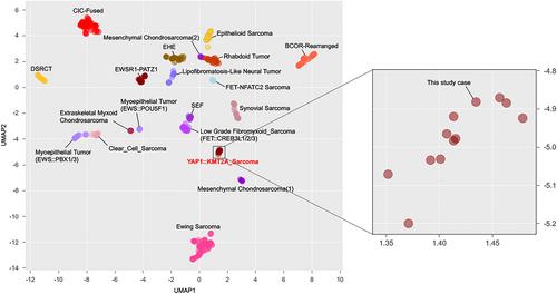
{"title":"YAP1:: kmt2a重排肉瘤:新病例报告,具有异常的形态和免疫组织化学特征","authors":"Caterina Fumagalli, Ruth Orellana, Sílvia Bagué, Malena Ferré, Allan Gonzalez, Lluis Catasús, Jaume Llauger, Ana Peiró, Paul Zamora Alarcón, Katarina Majercakova, Raúl Terés, Marie Karanian-Philippe, Franck Tirode, Cristina R. Antonescu","doi":"10.1002/gcc.70059","DOIUrl":null,"url":null,"abstract":"<p>Recurrent <i>KMT2A</i> and <i>YAP1</i> related fusions have recently been reported in various mesenchymal neoplasms of different histogenesis. First, <i>YAP1::KMT2A</i> fusions have been described in a subset of MUC4-negative sclerosing epithelioid fibrosarcomas (SEF), while <i>VIM::KMT2A</i> fusions in a handful of cases associated with an undifferentiated spindle cell phenotype lacking stromal hyalinization. On the other hand, <i>YAP1</i> gene rearrangements have been reported in a wide spectrum of sarcomas, including vascular neoplasms such as epithelioid hemangioendothelioma (EHE). Despite these molecular advances, occasional challenges in classification may occur even if the pathognomonic fusion is identified. In this study, we report such a case of a soft tissue sarcoma displaying an unusual morphology and immunoprofile, which remained unclassified even after a <i>YAP1::KMT2A</i> fusion was detected. The lesion occurred in the left leg of a 65-year-old female and microscopically closely resembled a SEF, with epithelioid morphology organized in cords, nests, and sheets in a heavy hyalinized background. Focally, the cells showed cytoplasmic vacuoles with eosinophilic material, reminiscent of the “blisters cells” seen in EHE. Moreover, by immunohistochemistry (IHC), the tumor showed diffuse reactivity for vascular markers, including ERG, CD31, CD34, and D2-40, as well as for TFE3, while being negative for MUC4, CAMTA1, smooth-muscle actin, desmin, S100 and keratins. Targeted RNA sequencing revealed a <i>YAP1::KMT2A</i> fusion. Based on this molecular result and the conflicting morphologic and IHC findings, a definitive distinction between a MUC4-negative SEF and an EHE could not been established. To further subclassify the lesion, subsequent clustering analysis using RNAseq signature was performed against a vast group of sarcoma types on the same array. Results showed that the tumor was in close proximity to the SEF group, admixed together with the other <i>YAP1::KMT2A</i> MUC4 negative SEF sarcomas. This case is highly instructive, as it shows another application of RNA sequencing in clinical practice when discordant or uncertain results between pathologic findings and fusion type may occur. Indeed, RNAseq signature could help, in this context, to better classify the tumor as a <i>YAP1::KMT2A</i> sarcoma instead of a vascular tumor. Larger series are needed to evaluate the pathogenesis of these tumors and the relevance of vascular markers expression.</p>","PeriodicalId":12700,"journal":{"name":"Genes, Chromosomes & Cancer","volume":"64 7","pages":""},"PeriodicalIF":2.8000,"publicationDate":"2025-07-11","publicationTypes":"Journal Article","fieldsOfStudy":null,"isOpenAccess":false,"openAccessPdf":"https://onlinelibrary.wiley.com/doi/epdf/10.1002/gcc.70059","citationCount":"0","resultStr":"{\"title\":\"YAP1::KMT2A-Rearranged Sarcoma: Report of a New Case With Unusual Morphology and Immunohistochemical Features\",\"authors\":\"Caterina Fumagalli, Ruth Orellana, Sílvia Bagué, Malena Ferré, Allan Gonzalez, Lluis Catasús, Jaume Llauger, Ana Peiró, Paul Zamora Alarcón, Katarina Majercakova, Raúl Terés, Marie Karanian-Philippe, Franck Tirode, Cristina R. Antonescu\",\"doi\":\"10.1002/gcc.70059\",\"DOIUrl\":null,\"url\":null,\"abstract\":\"<p>Recurrent <i>KMT2A</i> and <i>YAP1</i> related fusions have recently been reported in various mesenchymal neoplasms of different histogenesis. First, <i>YAP1::KMT2A</i> fusions have been described in a subset of MUC4-negative sclerosing epithelioid fibrosarcomas (SEF), while <i>VIM::KMT2A</i> fusions in a handful of cases associated with an undifferentiated spindle cell phenotype lacking stromal hyalinization. On the other hand, <i>YAP1</i> gene rearrangements have been reported in a wide spectrum of sarcomas, including vascular neoplasms such as epithelioid hemangioendothelioma (EHE). Despite these molecular advances, occasional challenges in classification may occur even if the pathognomonic fusion is identified. In this study, we report such a case of a soft tissue sarcoma displaying an unusual morphology and immunoprofile, which remained unclassified even after a <i>YAP1::KMT2A</i> fusion was detected. The lesion occurred in the left leg of a 65-year-old female and microscopically closely resembled a SEF, with epithelioid morphology organized in cords, nests, and sheets in a heavy hyalinized background. Focally, the cells showed cytoplasmic vacuoles with eosinophilic material, reminiscent of the “blisters cells” seen in EHE. Moreover, by immunohistochemistry (IHC), the tumor showed diffuse reactivity for vascular markers, including ERG, CD31, CD34, and D2-40, as well as for TFE3, while being negative for MUC4, CAMTA1, smooth-muscle actin, desmin, S100 and keratins. Targeted RNA sequencing revealed a <i>YAP1::KMT2A</i> fusion. Based on this molecular result and the conflicting morphologic and IHC findings, a definitive distinction between a MUC4-negative SEF and an EHE could not been established. To further subclassify the lesion, subsequent clustering analysis using RNAseq signature was performed against a vast group of sarcoma types on the same array. Results showed that the tumor was in close proximity to the SEF group, admixed together with the other <i>YAP1::KMT2A</i> MUC4 negative SEF sarcomas. This case is highly instructive, as it shows another application of RNA sequencing in clinical practice when discordant or uncertain results between pathologic findings and fusion type may occur. Indeed, RNAseq signature could help, in this context, to better classify the tumor as a <i>YAP1::KMT2A</i> sarcoma instead of a vascular tumor. Larger series are needed to evaluate the pathogenesis of these tumors and the relevance of vascular markers expression.</p>\",\"PeriodicalId\":12700,\"journal\":{\"name\":\"Genes, Chromosomes & Cancer\",\"volume\":\"64 7\",\"pages\":\"\"},\"PeriodicalIF\":2.8000,\"publicationDate\":\"2025-07-11\",\"publicationTypes\":\"Journal Article\",\"fieldsOfStudy\":null,\"isOpenAccess\":false,\"openAccessPdf\":\"https://onlinelibrary.wiley.com/doi/epdf/10.1002/gcc.70059\",\"citationCount\":\"0\",\"resultStr\":null,\"platform\":\"Semanticscholar\",\"paperid\":null,\"PeriodicalName\":\"Genes, Chromosomes & Cancer\",\"FirstCategoryId\":\"3\",\"ListUrlMain\":\"https://onlinelibrary.wiley.com/doi/10.1002/gcc.70059\",\"RegionNum\":2,\"RegionCategory\":\"医学\",\"ArticlePicture\":[],\"TitleCN\":null,\"AbstractTextCN\":null,\"PMCID\":null,\"EPubDate\":\"\",\"PubModel\":\"\",\"JCR\":\"Q2\",\"JCRName\":\"GENETICS & HEREDITY\",\"Score\":null,\"Total\":0}","platform":"Semanticscholar","paperid":null,"PeriodicalName":"Genes, Chromosomes & Cancer","FirstCategoryId":"3","ListUrlMain":"https://onlinelibrary.wiley.com/doi/10.1002/gcc.70059","RegionNum":2,"RegionCategory":"医学","ArticlePicture":[],"TitleCN":null,"AbstractTextCN":null,"PMCID":null,"EPubDate":"","PubModel":"","JCR":"Q2","JCRName":"GENETICS & HEREDITY","Score":null,"Total":0}
YAP1::KMT2A-Rearranged Sarcoma: Report of a New Case With Unusual Morphology and Immunohistochemical Features
Recurrent KMT2A and YAP1 related fusions have recently been reported in various mesenchymal neoplasms of different histogenesis. First, YAP1::KMT2A fusions have been described in a subset of MUC4-negative sclerosing epithelioid fibrosarcomas (SEF), while VIM::KMT2A fusions in a handful of cases associated with an undifferentiated spindle cell phenotype lacking stromal hyalinization. On the other hand, YAP1 gene rearrangements have been reported in a wide spectrum of sarcomas, including vascular neoplasms such as epithelioid hemangioendothelioma (EHE). Despite these molecular advances, occasional challenges in classification may occur even if the pathognomonic fusion is identified. In this study, we report such a case of a soft tissue sarcoma displaying an unusual morphology and immunoprofile, which remained unclassified even after a YAP1::KMT2A fusion was detected. The lesion occurred in the left leg of a 65-year-old female and microscopically closely resembled a SEF, with epithelioid morphology organized in cords, nests, and sheets in a heavy hyalinized background. Focally, the cells showed cytoplasmic vacuoles with eosinophilic material, reminiscent of the “blisters cells” seen in EHE. Moreover, by immunohistochemistry (IHC), the tumor showed diffuse reactivity for vascular markers, including ERG, CD31, CD34, and D2-40, as well as for TFE3, while being negative for MUC4, CAMTA1, smooth-muscle actin, desmin, S100 and keratins. Targeted RNA sequencing revealed a YAP1::KMT2A fusion. Based on this molecular result and the conflicting morphologic and IHC findings, a definitive distinction between a MUC4-negative SEF and an EHE could not been established. To further subclassify the lesion, subsequent clustering analysis using RNAseq signature was performed against a vast group of sarcoma types on the same array. Results showed that the tumor was in close proximity to the SEF group, admixed together with the other YAP1::KMT2A MUC4 negative SEF sarcomas. This case is highly instructive, as it shows another application of RNA sequencing in clinical practice when discordant or uncertain results between pathologic findings and fusion type may occur. Indeed, RNAseq signature could help, in this context, to better classify the tumor as a YAP1::KMT2A sarcoma instead of a vascular tumor. Larger series are needed to evaluate the pathogenesis of these tumors and the relevance of vascular markers expression.
期刊介绍:
Genes, Chromosomes & Cancer will offer rapid publication of original full-length research articles, perspectives, reviews and letters to the editors on genetic analysis as related to the study of neoplasia. The main scope of the journal is to communicate new insights into the etiology and/or pathogenesis of neoplasia, as well as molecular and cellular findings of relevance for the management of cancer patients. While preference will be given to research utilizing analytical and functional approaches, descriptive studies and case reports will also be welcomed when they offer insights regarding basic biological mechanisms or the clinical management of neoplastic disorders.
 求助内容:
求助内容: 应助结果提醒方式:


