{"title":"急性阑尾炎伴和不伴阑尾结石的CT评分和实验室参数的评价。","authors":"Tuğberk Baştürk, Mehmet Duran, Seda Baştürk","doi":"10.14744/tjtes.2005.75502","DOIUrl":null,"url":null,"abstract":"<p><strong>Background: </strong>With the growing interest in the non-operative management of acute appendicitis (AA), accurate diagnosis has become increasingly important. This study aimed to evaluate the computed tomography appendicitis score (CTAS), complete blood count (CBC), C-reactive protein (CRP), and systemic immune-inflammation index (SII) in patients diagnosed with AA with and without computed tomography-detected (CT-detected) appendicoliths. Additionally, the study compared these findings between patients with perforated and non-perforated appendicitis.</p><p><strong>Methods: </strong>Between January 2020 and January 2023, 294 patients diagnosed with AA were retrospectively analyzed. Of these, 140 (47.6%) had appendicoliths (Group 1), and 154 (52.4%) did not (Group 2). CT findings of AA and CTAS were evaluated. CBC parameters, CRP levels, and SII scores were compared between the groups, and the presence of appendix perforation was analyzed.</p><p><strong>Results: </strong>The mean diameter and wall thickness of the appendix, presence of intra-abdominal fluid, and severity of periappendi-ceal fat stranding were higher in Group 1 (p<0.001, p=0.024, p=0.009, p<0.001, respectively). The CTAS was also higher in Group 1 (7.51±2.35) compared to Group 2 (6.38±2.41; p<0.001). There was a positive correlation between the diameter of the appendicolith and CTAS (rho=0.450, p<0.001). In Group 1, CTAS was higher in patients with more than one appendicolith (p=0.003). Perforation was observed in 15 patients (10.7%) in Group 1 and five patients (3.2%) in Group 2, with a higher incidence in Group 1 (p=0.011). Among Group 1 patients, the perforation rate was higher in those with more than one appendicolith (p=0.019). The mean CTAS was higher in patients with appendiceal perforation (10±1.13) compared to those without perforation (7.22±2.29) (p<0.001). Monocyte (MONO) counts were also higher in Group 1 (p=0.002). Other CBC parameters, CRP levels, and SII scores did not differ significantly between Groups 1 and 2 (p>0.05). However, CRP levels and MONO counts were elevated in patients with perforated appendicitis (p<0.001 and p=0.026, respectively).</p><p><strong>Conclusion: </strong>Acute appendicitis with appendicoliths is associated with more pronounced inflammation and a higher rate of perforation. CTAS, CRP, and MONO levels tended to be elevated in cases of appendiceal perforation. A comprehensive evaluation incorporating the presence of appendicoliths, CTAS, and laboratory parameters may provide valuable insights into the severity of inflammation in AA.</p>","PeriodicalId":94263,"journal":{"name":"Ulusal travma ve acil cerrahi dergisi = Turkish journal of trauma & emergency surgery : TJTES","volume":"31 7","pages":"651-660"},"PeriodicalIF":1.0000,"publicationDate":"2025-07-01","publicationTypes":"Journal Article","fieldsOfStudy":null,"isOpenAccess":false,"openAccessPdf":"https://www.ncbi.nlm.nih.gov/pmc/articles/PMC12256970/pdf/","citationCount":"0","resultStr":"{\"title\":\"Evaluation of computed tomography (CT) appendicitis score and laboratory parameters in acute appendicitis with and without CT-detected appendicolith.\",\"authors\":\"Tuğberk Baştürk, Mehmet Duran, Seda Baştürk\",\"doi\":\"10.14744/tjtes.2005.75502\",\"DOIUrl\":null,\"url\":null,\"abstract\":\"<p><strong>Background: </strong>With the growing interest in the non-operative management of acute appendicitis (AA), accurate diagnosis has become increasingly important. This study aimed to evaluate the computed tomography appendicitis score (CTAS), complete blood count (CBC), C-reactive protein (CRP), and systemic immune-inflammation index (SII) in patients diagnosed with AA with and without computed tomography-detected (CT-detected) appendicoliths. Additionally, the study compared these findings between patients with perforated and non-perforated appendicitis.</p><p><strong>Methods: </strong>Between January 2020 and January 2023, 294 patients diagnosed with AA were retrospectively analyzed. Of these, 140 (47.6%) had appendicoliths (Group 1), and 154 (52.4%) did not (Group 2). CT findings of AA and CTAS were evaluated. CBC parameters, CRP levels, and SII scores were compared between the groups, and the presence of appendix perforation was analyzed.</p><p><strong>Results: </strong>The mean diameter and wall thickness of the appendix, presence of intra-abdominal fluid, and severity of periappendi-ceal fat stranding were higher in Group 1 (p<0.001, p=0.024, p=0.009, p<0.001, respectively). The CTAS was also higher in Group 1 (7.51±2.35) compared to Group 2 (6.38±2.41; p<0.001). There was a positive correlation between the diameter of the appendicolith and CTAS (rho=0.450, p<0.001). In Group 1, CTAS was higher in patients with more than one appendicolith (p=0.003). Perforation was observed in 15 patients (10.7%) in Group 1 and five patients (3.2%) in Group 2, with a higher incidence in Group 1 (p=0.011). Among Group 1 patients, the perforation rate was higher in those with more than one appendicolith (p=0.019). The mean CTAS was higher in patients with appendiceal perforation (10±1.13) compared to those without perforation (7.22±2.29) (p<0.001). Monocyte (MONO) counts were also higher in Group 1 (p=0.002). Other CBC parameters, CRP levels, and SII scores did not differ significantly between Groups 1 and 2 (p>0.05). However, CRP levels and MONO counts were elevated in patients with perforated appendicitis (p<0.001 and p=0.026, respectively).</p><p><strong>Conclusion: </strong>Acute appendicitis with appendicoliths is associated with more pronounced inflammation and a higher rate of perforation. CTAS, CRP, and MONO levels tended to be elevated in cases of appendiceal perforation. A comprehensive evaluation incorporating the presence of appendicoliths, CTAS, and laboratory parameters may provide valuable insights into the severity of inflammation in AA.</p>\",\"PeriodicalId\":94263,\"journal\":{\"name\":\"Ulusal travma ve acil cerrahi dergisi = Turkish journal of trauma & emergency surgery : TJTES\",\"volume\":\"31 7\",\"pages\":\"651-660\"},\"PeriodicalIF\":1.0000,\"publicationDate\":\"2025-07-01\",\"publicationTypes\":\"Journal Article\",\"fieldsOfStudy\":null,\"isOpenAccess\":false,\"openAccessPdf\":\"https://www.ncbi.nlm.nih.gov/pmc/articles/PMC12256970/pdf/\",\"citationCount\":\"0\",\"resultStr\":null,\"platform\":\"Semanticscholar\",\"paperid\":null,\"PeriodicalName\":\"Ulusal travma ve acil cerrahi dergisi = Turkish journal of trauma & emergency surgery : TJTES\",\"FirstCategoryId\":\"1085\",\"ListUrlMain\":\"https://doi.org/10.14744/tjtes.2005.75502\",\"RegionNum\":0,\"RegionCategory\":null,\"ArticlePicture\":[],\"TitleCN\":null,\"AbstractTextCN\":null,\"PMCID\":null,\"EPubDate\":\"\",\"PubModel\":\"\",\"JCR\":\"\",\"JCRName\":\"\",\"Score\":null,\"Total\":0}","platform":"Semanticscholar","paperid":null,"PeriodicalName":"Ulusal travma ve acil cerrahi dergisi = Turkish journal of trauma & emergency surgery : TJTES","FirstCategoryId":"1085","ListUrlMain":"https://doi.org/10.14744/tjtes.2005.75502","RegionNum":0,"RegionCategory":null,"ArticlePicture":[],"TitleCN":null,"AbstractTextCN":null,"PMCID":null,"EPubDate":"","PubModel":"","JCR":"","JCRName":"","Score":null,"Total":0}
Evaluation of computed tomography (CT) appendicitis score and laboratory parameters in acute appendicitis with and without CT-detected appendicolith.
Background: With the growing interest in the non-operative management of acute appendicitis (AA), accurate diagnosis has become increasingly important. This study aimed to evaluate the computed tomography appendicitis score (CTAS), complete blood count (CBC), C-reactive protein (CRP), and systemic immune-inflammation index (SII) in patients diagnosed with AA with and without computed tomography-detected (CT-detected) appendicoliths. Additionally, the study compared these findings between patients with perforated and non-perforated appendicitis.
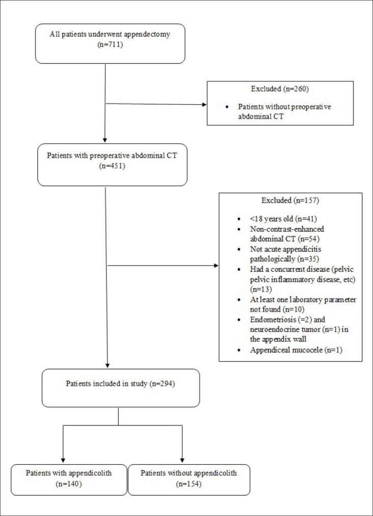
Methods: Between January 2020 and January 2023, 294 patients diagnosed with AA were retrospectively analyzed. Of these, 140 (47.6%) had appendicoliths (Group 1), and 154 (52.4%) did not (Group 2). CT findings of AA and CTAS were evaluated. CBC parameters, CRP levels, and SII scores were compared between the groups, and the presence of appendix perforation was analyzed.
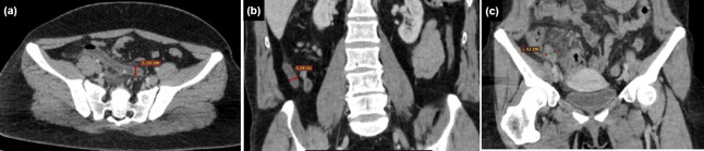
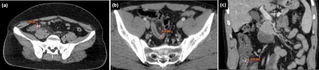
Results: The mean diameter and wall thickness of the appendix, presence of intra-abdominal fluid, and severity of periappendi-ceal fat stranding were higher in Group 1 (p<0.001, p=0.024, p=0.009, p<0.001, respectively). The CTAS was also higher in Group 1 (7.51±2.35) compared to Group 2 (6.38±2.41; p<0.001). There was a positive correlation between the diameter of the appendicolith and CTAS (rho=0.450, p<0.001). In Group 1, CTAS was higher in patients with more than one appendicolith (p=0.003). Perforation was observed in 15 patients (10.7%) in Group 1 and five patients (3.2%) in Group 2, with a higher incidence in Group 1 (p=0.011). Among Group 1 patients, the perforation rate was higher in those with more than one appendicolith (p=0.019). The mean CTAS was higher in patients with appendiceal perforation (10±1.13) compared to those without perforation (7.22±2.29) (p<0.001). Monocyte (MONO) counts were also higher in Group 1 (p=0.002). Other CBC parameters, CRP levels, and SII scores did not differ significantly between Groups 1 and 2 (p>0.05). However, CRP levels and MONO counts were elevated in patients with perforated appendicitis (p<0.001 and p=0.026, respectively).
Conclusion: Acute appendicitis with appendicoliths is associated with more pronounced inflammation and a higher rate of perforation. CTAS, CRP, and MONO levels tended to be elevated in cases of appendiceal perforation. A comprehensive evaluation incorporating the presence of appendicoliths, CTAS, and laboratory parameters may provide valuable insights into the severity of inflammation in AA.

 求助内容:
求助内容: 应助结果提醒方式:


