Cristian Aletto, Martina Marsiolo, Michela Florio, Angelo Gabriele Aulisa, Renato Maria Toniolo, Francesco Falciglia, Nicola Maffulli
{"title":"从分类到手术计划,计算机断层扫描在儿童踝关节骨折中的应用。","authors":"Cristian Aletto, Martina Marsiolo, Michela Florio, Angelo Gabriele Aulisa, Renato Maria Toniolo, Francesco Falciglia, Nicola Maffulli","doi":"10.1186/s10195-025-00840-3","DOIUrl":null,"url":null,"abstract":"<p><strong>Background: </strong>Ankle fractures are common in the pediatric population. Plain radiographs provide sufficient information for the diagnosis, but computed tomography (CT) can help to study the configuration of fracture and to plan fixation. Our study aims to study pediatric population with ankle fracture, understanding whether CT scans should be extended to all ankle fractures admitted to the Orthopaedic Department after a first radiographic evaluation, independent of the pattern of physeal plate fracture.</p><p><strong>Materials and methods: </strong>Data about patients with ankle fractures admitted to the Orthopaedic Department were retrieved. The diagnosis and classification of ankle fractures obtained from plain radiographs were compared with those obtained from CT scans. For each patient, data about conservative or surgical management were retrieved. After collecting all the mentioned data, a survey with 61 plain radiographs of children's ankle fractures was proposed to 16 orthopedic surgeons of the department divided into three groups according to their years of experience in Paediatric Orthopaedics and Trauma. The survey consisted of five questions for each radiograph regarding Salter-Harris (SH) classification, management, indication for CT, number, and direction of screws (if needed).</p><p><strong>Results: </strong>A total of 130 patients with ankle fractures satisfied the inclusion criteria and only 26 of them were classified according to the SH classification by orthopedic surgeons or radiologists after plain radiography. Almost all pediatric patients with ankle fractures admitted to the Orthopaedic Department, after evaluation of plain radiographs in the emergency department (ED), underwent CT with three-dimensional (3D) reconstruction to plan fixation or nonoperative management. CT may lead to reclassification of some fractures, showing that SHIV fractures may be more common than expected. A total of 6 orthopedic surgeons answered the survey on 61 ankle fracture plain radiographs. Independent of their experience, orthopedic surgeons tend to respond similarly to SH classification and fracture management, while they have contrasting opinions about performing CT scans. Analyzing their response to the number of screws, entry points, and directions and comparing them with postoperation radiographs, the results between responders were very discordant.</p><p><strong>Conclusion: </strong>In children's ankle fracture involving the physeal plate, the SH classification, fracture management planning, the identification of the entry point and the direction of the screw could be more accurate using CT compared with plain radiographs.</p><p><strong>Levels of evidence: </strong>Level IV, according to the Oxford 2011 Levels of Evidence.</p>","PeriodicalId":48603,"journal":{"name":"Journal of Orthopaedics and Traumatology","volume":"26 1","pages":"45"},"PeriodicalIF":3.7000,"publicationDate":"2025-07-08","publicationTypes":"Journal Article","fieldsOfStudy":null,"isOpenAccess":false,"openAccessPdf":"https://www.ncbi.nlm.nih.gov/pmc/articles/PMC12237838/pdf/","citationCount":"0","resultStr":"{\"title\":\"Utility of computed tomography in children's ankle fractures from classification to surgical planning.\",\"authors\":\"Cristian Aletto, Martina Marsiolo, Michela Florio, Angelo Gabriele Aulisa, Renato Maria Toniolo, Francesco Falciglia, Nicola Maffulli\",\"doi\":\"10.1186/s10195-025-00840-3\",\"DOIUrl\":null,\"url\":null,\"abstract\":\"<p><strong>Background: </strong>Ankle fractures are common in the pediatric population. Plain radiographs provide sufficient information for the diagnosis, but computed tomography (CT) can help to study the configuration of fracture and to plan fixation. Our study aims to study pediatric population with ankle fracture, understanding whether CT scans should be extended to all ankle fractures admitted to the Orthopaedic Department after a first radiographic evaluation, independent of the pattern of physeal plate fracture.</p><p><strong>Materials and methods: </strong>Data about patients with ankle fractures admitted to the Orthopaedic Department were retrieved. The diagnosis and classification of ankle fractures obtained from plain radiographs were compared with those obtained from CT scans. For each patient, data about conservative or surgical management were retrieved. After collecting all the mentioned data, a survey with 61 plain radiographs of children's ankle fractures was proposed to 16 orthopedic surgeons of the department divided into three groups according to their years of experience in Paediatric Orthopaedics and Trauma. The survey consisted of five questions for each radiograph regarding Salter-Harris (SH) classification, management, indication for CT, number, and direction of screws (if needed).</p><p><strong>Results: </strong>A total of 130 patients with ankle fractures satisfied the inclusion criteria and only 26 of them were classified according to the SH classification by orthopedic surgeons or radiologists after plain radiography. Almost all pediatric patients with ankle fractures admitted to the Orthopaedic Department, after evaluation of plain radiographs in the emergency department (ED), underwent CT with three-dimensional (3D) reconstruction to plan fixation or nonoperative management. CT may lead to reclassification of some fractures, showing that SHIV fractures may be more common than expected. A total of 6 orthopedic surgeons answered the survey on 61 ankle fracture plain radiographs. Independent of their experience, orthopedic surgeons tend to respond similarly to SH classification and fracture management, while they have contrasting opinions about performing CT scans. Analyzing their response to the number of screws, entry points, and directions and comparing them with postoperation radiographs, the results between responders were very discordant.</p><p><strong>Conclusion: </strong>In children's ankle fracture involving the physeal plate, the SH classification, fracture management planning, the identification of the entry point and the direction of the screw could be more accurate using CT compared with plain radiographs.</p><p><strong>Levels of evidence: </strong>Level IV, according to the Oxford 2011 Levels of Evidence.</p>\",\"PeriodicalId\":48603,\"journal\":{\"name\":\"Journal of Orthopaedics and Traumatology\",\"volume\":\"26 1\",\"pages\":\"45\"},\"PeriodicalIF\":3.7000,\"publicationDate\":\"2025-07-08\",\"publicationTypes\":\"Journal Article\",\"fieldsOfStudy\":null,\"isOpenAccess\":false,\"openAccessPdf\":\"https://www.ncbi.nlm.nih.gov/pmc/articles/PMC12237838/pdf/\",\"citationCount\":\"0\",\"resultStr\":null,\"platform\":\"Semanticscholar\",\"paperid\":null,\"PeriodicalName\":\"Journal of Orthopaedics and Traumatology\",\"FirstCategoryId\":\"3\",\"ListUrlMain\":\"https://doi.org/10.1186/s10195-025-00840-3\",\"RegionNum\":2,\"RegionCategory\":\"医学\",\"ArticlePicture\":[],\"TitleCN\":null,\"AbstractTextCN\":null,\"PMCID\":null,\"EPubDate\":\"\",\"PubModel\":\"\",\"JCR\":\"Q1\",\"JCRName\":\"ORTHOPEDICS\",\"Score\":null,\"Total\":0}","platform":"Semanticscholar","paperid":null,"PeriodicalName":"Journal of Orthopaedics and Traumatology","FirstCategoryId":"3","ListUrlMain":"https://doi.org/10.1186/s10195-025-00840-3","RegionNum":2,"RegionCategory":"医学","ArticlePicture":[],"TitleCN":null,"AbstractTextCN":null,"PMCID":null,"EPubDate":"","PubModel":"","JCR":"Q1","JCRName":"ORTHOPEDICS","Score":null,"Total":0}
Utility of computed tomography in children's ankle fractures from classification to surgical planning.
Background: Ankle fractures are common in the pediatric population. Plain radiographs provide sufficient information for the diagnosis, but computed tomography (CT) can help to study the configuration of fracture and to plan fixation. Our study aims to study pediatric population with ankle fracture, understanding whether CT scans should be extended to all ankle fractures admitted to the Orthopaedic Department after a first radiographic evaluation, independent of the pattern of physeal plate fracture.
Materials and methods: Data about patients with ankle fractures admitted to the Orthopaedic Department were retrieved. The diagnosis and classification of ankle fractures obtained from plain radiographs were compared with those obtained from CT scans. For each patient, data about conservative or surgical management were retrieved. After collecting all the mentioned data, a survey with 61 plain radiographs of children's ankle fractures was proposed to 16 orthopedic surgeons of the department divided into three groups according to their years of experience in Paediatric Orthopaedics and Trauma. The survey consisted of five questions for each radiograph regarding Salter-Harris (SH) classification, management, indication for CT, number, and direction of screws (if needed).
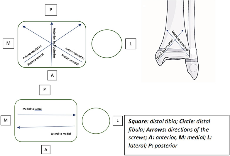
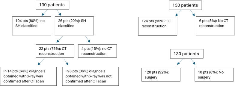
Results: A total of 130 patients with ankle fractures satisfied the inclusion criteria and only 26 of them were classified according to the SH classification by orthopedic surgeons or radiologists after plain radiography. Almost all pediatric patients with ankle fractures admitted to the Orthopaedic Department, after evaluation of plain radiographs in the emergency department (ED), underwent CT with three-dimensional (3D) reconstruction to plan fixation or nonoperative management. CT may lead to reclassification of some fractures, showing that SHIV fractures may be more common than expected. A total of 6 orthopedic surgeons answered the survey on 61 ankle fracture plain radiographs. Independent of their experience, orthopedic surgeons tend to respond similarly to SH classification and fracture management, while they have contrasting opinions about performing CT scans. Analyzing their response to the number of screws, entry points, and directions and comparing them with postoperation radiographs, the results between responders were very discordant.
Conclusion: In children's ankle fracture involving the physeal plate, the SH classification, fracture management planning, the identification of the entry point and the direction of the screw could be more accurate using CT compared with plain radiographs.
Levels of evidence: Level IV, according to the Oxford 2011 Levels of Evidence.
期刊介绍:
The Journal of Orthopaedics and Traumatology, the official open access peer-reviewed journal of the Italian Society of Orthopaedics and Traumatology, publishes original papers reporting basic or clinical research in the field of orthopaedic and traumatologic surgery, as well as systematic reviews, brief communications, case reports and letters to the Editor. Narrative instructional reviews and commentaries to original articles may be commissioned by Editors from eminent colleagues. The Journal of Orthopaedics and Traumatology aims to be an international forum for the communication and exchange of ideas concerning the various aspects of orthopaedics and musculoskeletal trauma.

 求助内容:
求助内容: 应助结果提醒方式:


