Molecular VisionPub Date : 2025-05-17eCollection Date: 2025-01-01
Mangesh Bawankar, Bhaswati Sengupta, Sujata Malik, Pratik Sen, Ashwani K Thakur
{"title":"利用荧光相关光谱揭示γ - d -晶体蛋白聚集途径以了解白内障的形成。","authors":"Mangesh Bawankar, Bhaswati Sengupta, Sujata Malik, Pratik Sen, Ashwani K Thakur","doi":"","DOIUrl":null,"url":null,"abstract":"<p><strong>Purpose: </strong>To characterize the aggregation behavior of the γD-crystallin protein in an acidic environment with a focus on the formation of intermediate species. The research employs fluorescence correlation spectroscopy to unravel the intricate molecular events leading to aggregation, contributing to a comprehensive understanding of cataract formation.</p><p><strong>Methods: </strong>The kinetics of γD-crystallin protein aggregation were studied with a reversed-phase high-performance liquid chromatography sedimentation assay, a ThT binding assay, and light scattering. We used fluorescence correlation spectroscopy (FCS) to recognize intermediate aggregate species and characterized them with Fourier transform infrared spectroscopy (FTIR). Further, the morphologic characterization of aggregates was done by transmission electron microscopy (TEM), and their hydrophobic characteristics were analyzed using the 8-anilino-1-naphthalenesulfonic acid binding assay.</p><p><strong>Results: </strong>A negligible lag phase was observed in the aggregation kinetic experiments of the γD-crystallin protein. Pentamer, 25-mer, and higher oligomer intermediates were formed on the aggregation pathway. Conformation studies by FCS and FTIR have shown that oligomers are rich in cross-β sheet and random coil structure; however, they constitute more α-helix and less cross-β sheet structure than fibrils. TEM analysis revealed the approximate size of oligomers (diameter ~10 nm), protofibrils (~15 nm), and fibrils (~15 to ~35 nm).</p><p><strong>Conclusions: </strong>In this study, we reported the presence of various intermediate aggregate species formed on the aggregation pathway of γD-crystallin protein at low pH. This will open new areas of research in understanding the detailed aggregation mechanism and aggregation hotspot within unfolded γD-crystallin monomers. The insights gained will also pave the way for future research in the realm of amyloid formation in cataract.</p>","PeriodicalId":18866,"journal":{"name":"Molecular Vision","volume":"31 ","pages":"190-202"},"PeriodicalIF":1.4000,"publicationDate":"2025-05-17","publicationTypes":"Journal Article","fieldsOfStudy":null,"isOpenAccess":false,"openAccessPdf":"https://www.ncbi.nlm.nih.gov/pmc/articles/PMC12221310/pdf/","citationCount":"0","resultStr":"{\"title\":\"Unravelling γD-crystallin aggregation pathway to understand cataract formation using fluorescence correlation spectroscopy.\",\"authors\":\"Mangesh Bawankar, Bhaswati Sengupta, Sujata Malik, Pratik Sen, Ashwani K Thakur\",\"doi\":\"\",\"DOIUrl\":null,\"url\":null,\"abstract\":\"<p><strong>Purpose: </strong>To characterize the aggregation behavior of the γD-crystallin protein in an acidic environment with a focus on the formation of intermediate species. The research employs fluorescence correlation spectroscopy to unravel the intricate molecular events leading to aggregation, contributing to a comprehensive understanding of cataract formation.</p><p><strong>Methods: </strong>The kinetics of γD-crystallin protein aggregation were studied with a reversed-phase high-performance liquid chromatography sedimentation assay, a ThT binding assay, and light scattering. We used fluorescence correlation spectroscopy (FCS) to recognize intermediate aggregate species and characterized them with Fourier transform infrared spectroscopy (FTIR). Further, the morphologic characterization of aggregates was done by transmission electron microscopy (TEM), and their hydrophobic characteristics were analyzed using the 8-anilino-1-naphthalenesulfonic acid binding assay.</p><p><strong>Results: </strong>A negligible lag phase was observed in the aggregation kinetic experiments of the γD-crystallin protein. Pentamer, 25-mer, and higher oligomer intermediates were formed on the aggregation pathway. Conformation studies by FCS and FTIR have shown that oligomers are rich in cross-β sheet and random coil structure; however, they constitute more α-helix and less cross-β sheet structure than fibrils. TEM analysis revealed the approximate size of oligomers (diameter ~10 nm), protofibrils (~15 nm), and fibrils (~15 to ~35 nm).</p><p><strong>Conclusions: </strong>In this study, we reported the presence of various intermediate aggregate species formed on the aggregation pathway of γD-crystallin protein at low pH. This will open new areas of research in understanding the detailed aggregation mechanism and aggregation hotspot within unfolded γD-crystallin monomers. The insights gained will also pave the way for future research in the realm of amyloid formation in cataract.</p>\",\"PeriodicalId\":18866,\"journal\":{\"name\":\"Molecular Vision\",\"volume\":\"31 \",\"pages\":\"190-202\"},\"PeriodicalIF\":1.4000,\"publicationDate\":\"2025-05-17\",\"publicationTypes\":\"Journal Article\",\"fieldsOfStudy\":null,\"isOpenAccess\":false,\"openAccessPdf\":\"https://www.ncbi.nlm.nih.gov/pmc/articles/PMC12221310/pdf/\",\"citationCount\":\"0\",\"resultStr\":null,\"platform\":\"Semanticscholar\",\"paperid\":null,\"PeriodicalName\":\"Molecular Vision\",\"FirstCategoryId\":\"3\",\"ListUrlMain\":\"\",\"RegionNum\":3,\"RegionCategory\":\"医学\",\"ArticlePicture\":[],\"TitleCN\":null,\"AbstractTextCN\":null,\"PMCID\":null,\"EPubDate\":\"2025/1/1 0:00:00\",\"PubModel\":\"eCollection\",\"JCR\":\"Q4\",\"JCRName\":\"BIOCHEMISTRY & MOLECULAR BIOLOGY\",\"Score\":null,\"Total\":0}","platform":"Semanticscholar","paperid":null,"PeriodicalName":"Molecular Vision","FirstCategoryId":"3","ListUrlMain":"","RegionNum":3,"RegionCategory":"医学","ArticlePicture":[],"TitleCN":null,"AbstractTextCN":null,"PMCID":null,"EPubDate":"2025/1/1 0:00:00","PubModel":"eCollection","JCR":"Q4","JCRName":"BIOCHEMISTRY & MOLECULAR BIOLOGY","Score":null,"Total":0}
引用次数: 0
摘要
目的:研究γ - d -结晶蛋白在酸性环境中的聚集行为,重点研究中间产物的形成。该研究采用荧光相关光谱来揭示导致聚集的复杂分子事件,有助于全面了解白内障的形成。方法:采用反相高效液相色谱沉淀法、ThT结合法和光散射法研究γ - d-晶体蛋白聚集动力学。利用荧光相关光谱(FCS)识别中间聚集体,并用傅里叶变换红外光谱(FTIR)对其进行表征。利用透射电子显微镜(TEM)对聚集体进行了形态表征,并利用8-苯胺-1-萘磺酸结合实验分析了聚集体的疏水特性。结果:在γ - d -晶体蛋白的聚集动力学实验中,观察到一个可以忽略不计的滞后期。聚集途径上形成了五聚体、25聚体和更高的低聚体中间体。FCS和FTIR的构象研究表明,低聚物具有丰富的交叉β片和随机线圈结构;但与原纤维相比,它们具有更多的α-螺旋结构和较少的交叉-β片结构。TEM分析显示了低聚物(直径~10 nm)、原纤维(~15 nm)和原纤维(~15 ~ ~35 nm)的大致尺寸。结论:在本研究中,我们报道了在低ph下γ d -结晶蛋白聚集途径上形成的多种中间聚集物质的存在,这将为了解未折叠的γ d -结晶蛋白单体内部的详细聚集机制和聚集热点开辟新的研究领域。所获得的见解也将为未来在白内障淀粉样蛋白形成领域的研究铺平道路。
Unravelling γD-crystallin aggregation pathway to understand cataract formation using fluorescence correlation spectroscopy.
Purpose: To characterize the aggregation behavior of the γD-crystallin protein in an acidic environment with a focus on the formation of intermediate species. The research employs fluorescence correlation spectroscopy to unravel the intricate molecular events leading to aggregation, contributing to a comprehensive understanding of cataract formation.
Methods: The kinetics of γD-crystallin protein aggregation were studied with a reversed-phase high-performance liquid chromatography sedimentation assay, a ThT binding assay, and light scattering. We used fluorescence correlation spectroscopy (FCS) to recognize intermediate aggregate species and characterized them with Fourier transform infrared spectroscopy (FTIR). Further, the morphologic characterization of aggregates was done by transmission electron microscopy (TEM), and their hydrophobic characteristics were analyzed using the 8-anilino-1-naphthalenesulfonic acid binding assay.
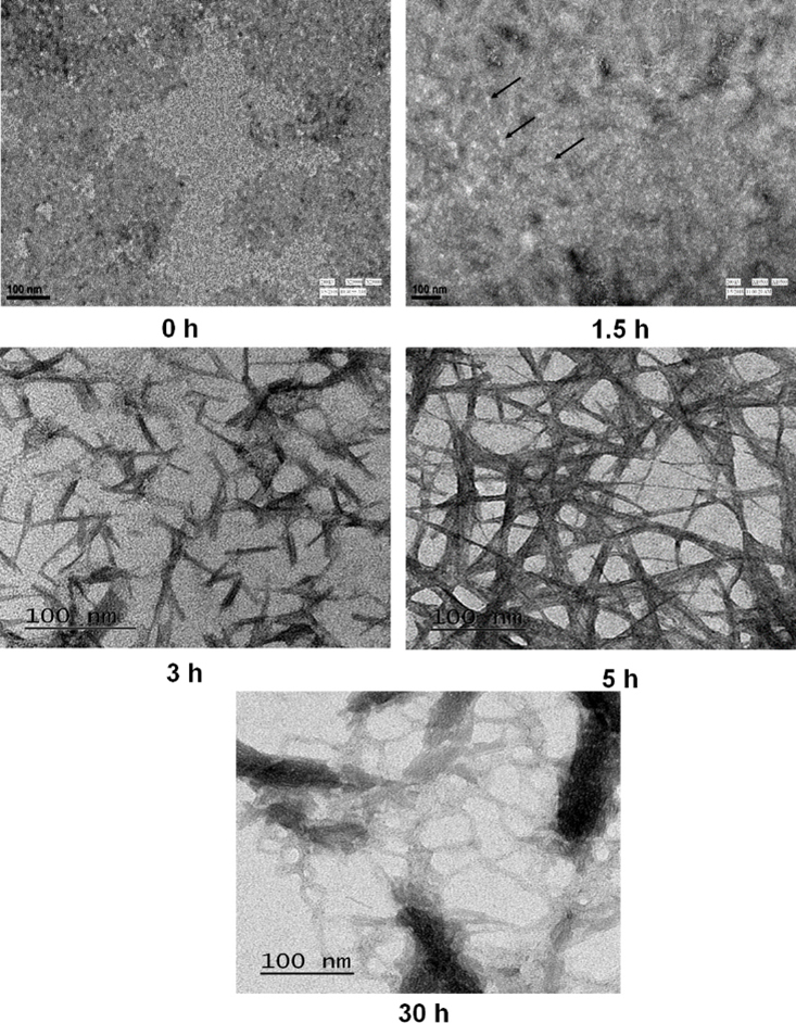
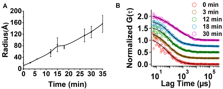
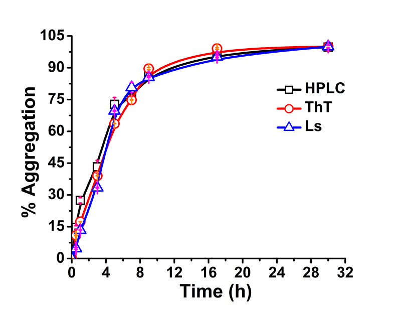
Results: A negligible lag phase was observed in the aggregation kinetic experiments of the γD-crystallin protein. Pentamer, 25-mer, and higher oligomer intermediates were formed on the aggregation pathway. Conformation studies by FCS and FTIR have shown that oligomers are rich in cross-β sheet and random coil structure; however, they constitute more α-helix and less cross-β sheet structure than fibrils. TEM analysis revealed the approximate size of oligomers (diameter ~10 nm), protofibrils (~15 nm), and fibrils (~15 to ~35 nm).
Conclusions: In this study, we reported the presence of various intermediate aggregate species formed on the aggregation pathway of γD-crystallin protein at low pH. This will open new areas of research in understanding the detailed aggregation mechanism and aggregation hotspot within unfolded γD-crystallin monomers. The insights gained will also pave the way for future research in the realm of amyloid formation in cataract.
期刊介绍:
Molecular Vision is a peer-reviewed journal dedicated to the dissemination of research results in molecular biology, cell biology, and the genetics of the visual system (ocular and cortical).
Molecular Vision publishes articles presenting original research that has not previously been published and comprehensive articles reviewing the current status of a particular field or topic. Submissions to Molecular Vision are subjected to rigorous peer review. Molecular Vision does NOT publish preprints.
For authors, Molecular Vision provides a rapid means of communicating important results. Access to Molecular Vision is free and unrestricted, allowing the widest possible audience for your article. Digital publishing allows you to use color images freely (and without fees). Additionally, you may publish animations, sounds, or other supplementary information that clarifies or supports your article. Each of the authors of an article may also list an electronic mail address (which will be updated upon request) to give interested readers easy access to authors.
 求助内容:
求助内容: 应助结果提醒方式:


