Yong Zeng, Jiaming Zhou, Yichao Li, Bruno Alvisio, Jacob Czech, David Bissig, Haohua Qian
{"title":"基于人工智能的OCT图像分割评价碘酸钠致视网膜变性视网膜结构变化。","authors":"Yong Zeng, Jiaming Zhou, Yichao Li, Bruno Alvisio, Jacob Czech, David Bissig, Haohua Qian","doi":"10.3389/fncel.2025.1605639","DOIUrl":null,"url":null,"abstract":"<p><p>Segmentations of retinal optical coherence tomography (OCT) images provide valuable information about each specific retinal layer. However, processing images from degenerative retina remains challenging. This study developed artificial intelligence (AI)-based segmentation to analyze structure changes in sodium iodate (SI)-treated mice. The software is capable of segmenting seven retinal layers and one choroid layer. Analyzing OCT images captured at days post SI-injection (PI) revealed early changes in the retinal pigment epithelium (RPE) layer, with increase in thickness and reduction in reflectance calculated by estimated Attenuation Coefficients (eAC). On the other hand, eAC for outer nuclear layer (ONL) exhibited early and sustained increase after SI treatment. SI induced exponential reduction in ONL thickness with a half-reduction time of about 3 days, indicating progressive photoreceptor degeneration. The extent of degeneration was correlated with ONL eAC level at PI1. Inner retinal layers showed bi-phasic reactions, with initial increases in layer thickness that peaked at around PI3, followed by gradual reduction to lower than baseline levels. In addition, SI also induced transient increases in vitreous particles concentrated around the optic nerve head. Furthermore, there was a gradual reduction of choroid thickness after SI treatment. These results indicate the AI-segmentation tool's usefulness for providing a sensitive and accurate assessment of structure changes in diseased retina and revealed more detailed characterization of SI-induced degeneration in all retinal layers with distinct time courses. Our results also support ONL reflectance changes as an early biomarker for retinal degeneration.</p>","PeriodicalId":12432,"journal":{"name":"Frontiers in Cellular Neuroscience","volume":"19 ","pages":"1605639"},"PeriodicalIF":4.0000,"publicationDate":"2025-06-18","publicationTypes":"Journal Article","fieldsOfStudy":null,"isOpenAccess":false,"openAccessPdf":"https://www.ncbi.nlm.nih.gov/pmc/articles/PMC12213626/pdf/","citationCount":"0","resultStr":"{\"title\":\"Evaluation of retinal structure changes with AI-based OCT image segmentation for sodium iodate induced retinal degeneration.\",\"authors\":\"Yong Zeng, Jiaming Zhou, Yichao Li, Bruno Alvisio, Jacob Czech, David Bissig, Haohua Qian\",\"doi\":\"10.3389/fncel.2025.1605639\",\"DOIUrl\":null,\"url\":null,\"abstract\":\"<p><p>Segmentations of retinal optical coherence tomography (OCT) images provide valuable information about each specific retinal layer. However, processing images from degenerative retina remains challenging. This study developed artificial intelligence (AI)-based segmentation to analyze structure changes in sodium iodate (SI)-treated mice. The software is capable of segmenting seven retinal layers and one choroid layer. Analyzing OCT images captured at days post SI-injection (PI) revealed early changes in the retinal pigment epithelium (RPE) layer, with increase in thickness and reduction in reflectance calculated by estimated Attenuation Coefficients (eAC). On the other hand, eAC for outer nuclear layer (ONL) exhibited early and sustained increase after SI treatment. SI induced exponential reduction in ONL thickness with a half-reduction time of about 3 days, indicating progressive photoreceptor degeneration. The extent of degeneration was correlated with ONL eAC level at PI1. Inner retinal layers showed bi-phasic reactions, with initial increases in layer thickness that peaked at around PI3, followed by gradual reduction to lower than baseline levels. In addition, SI also induced transient increases in vitreous particles concentrated around the optic nerve head. Furthermore, there was a gradual reduction of choroid thickness after SI treatment. These results indicate the AI-segmentation tool's usefulness for providing a sensitive and accurate assessment of structure changes in diseased retina and revealed more detailed characterization of SI-induced degeneration in all retinal layers with distinct time courses. Our results also support ONL reflectance changes as an early biomarker for retinal degeneration.</p>\",\"PeriodicalId\":12432,\"journal\":{\"name\":\"Frontiers in Cellular Neuroscience\",\"volume\":\"19 \",\"pages\":\"1605639\"},\"PeriodicalIF\":4.0000,\"publicationDate\":\"2025-06-18\",\"publicationTypes\":\"Journal Article\",\"fieldsOfStudy\":null,\"isOpenAccess\":false,\"openAccessPdf\":\"https://www.ncbi.nlm.nih.gov/pmc/articles/PMC12213626/pdf/\",\"citationCount\":\"0\",\"resultStr\":null,\"platform\":\"Semanticscholar\",\"paperid\":null,\"PeriodicalName\":\"Frontiers in Cellular Neuroscience\",\"FirstCategoryId\":\"3\",\"ListUrlMain\":\"https://doi.org/10.3389/fncel.2025.1605639\",\"RegionNum\":3,\"RegionCategory\":\"医学\",\"ArticlePicture\":[],\"TitleCN\":null,\"AbstractTextCN\":null,\"PMCID\":null,\"EPubDate\":\"2025/1/1 0:00:00\",\"PubModel\":\"eCollection\",\"JCR\":\"Q2\",\"JCRName\":\"NEUROSCIENCES\",\"Score\":null,\"Total\":0}","platform":"Semanticscholar","paperid":null,"PeriodicalName":"Frontiers in Cellular Neuroscience","FirstCategoryId":"3","ListUrlMain":"https://doi.org/10.3389/fncel.2025.1605639","RegionNum":3,"RegionCategory":"医学","ArticlePicture":[],"TitleCN":null,"AbstractTextCN":null,"PMCID":null,"EPubDate":"2025/1/1 0:00:00","PubModel":"eCollection","JCR":"Q2","JCRName":"NEUROSCIENCES","Score":null,"Total":0}
Evaluation of retinal structure changes with AI-based OCT image segmentation for sodium iodate induced retinal degeneration.
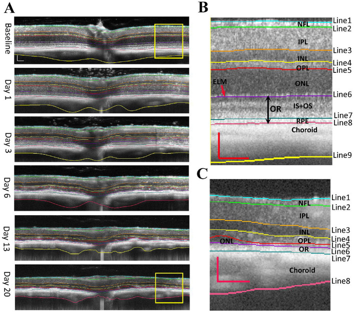
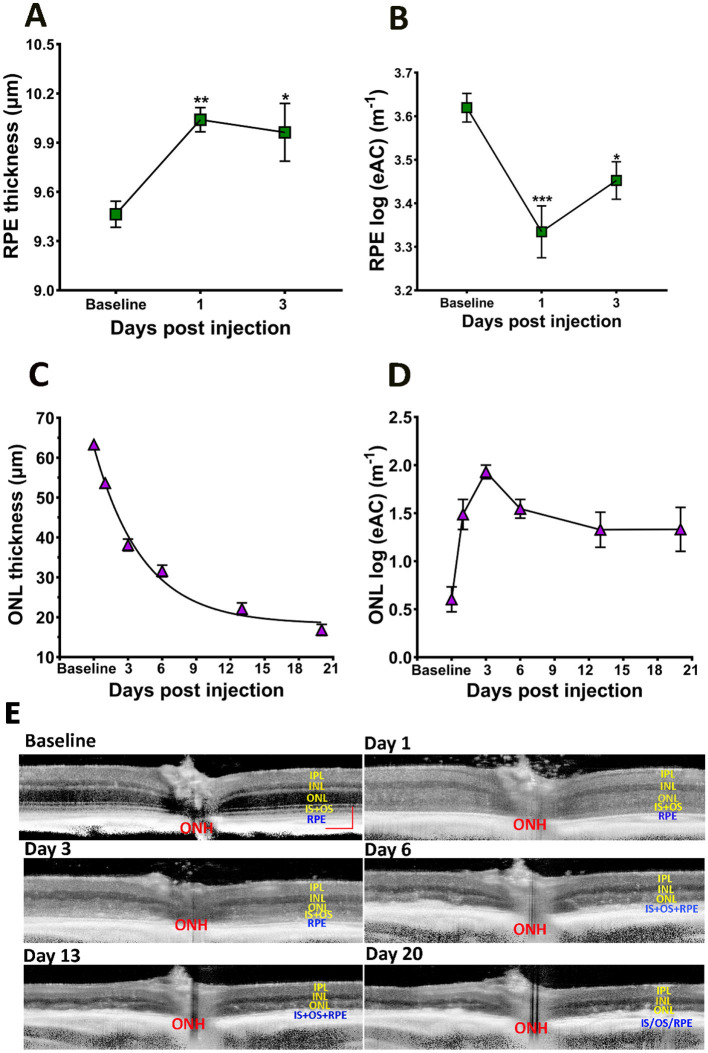
Segmentations of retinal optical coherence tomography (OCT) images provide valuable information about each specific retinal layer. However, processing images from degenerative retina remains challenging. This study developed artificial intelligence (AI)-based segmentation to analyze structure changes in sodium iodate (SI)-treated mice. The software is capable of segmenting seven retinal layers and one choroid layer. Analyzing OCT images captured at days post SI-injection (PI) revealed early changes in the retinal pigment epithelium (RPE) layer, with increase in thickness and reduction in reflectance calculated by estimated Attenuation Coefficients (eAC). On the other hand, eAC for outer nuclear layer (ONL) exhibited early and sustained increase after SI treatment. SI induced exponential reduction in ONL thickness with a half-reduction time of about 3 days, indicating progressive photoreceptor degeneration. The extent of degeneration was correlated with ONL eAC level at PI1. Inner retinal layers showed bi-phasic reactions, with initial increases in layer thickness that peaked at around PI3, followed by gradual reduction to lower than baseline levels. In addition, SI also induced transient increases in vitreous particles concentrated around the optic nerve head. Furthermore, there was a gradual reduction of choroid thickness after SI treatment. These results indicate the AI-segmentation tool's usefulness for providing a sensitive and accurate assessment of structure changes in diseased retina and revealed more detailed characterization of SI-induced degeneration in all retinal layers with distinct time courses. Our results also support ONL reflectance changes as an early biomarker for retinal degeneration.
期刊介绍:
Frontiers in Cellular Neuroscience is a leading journal in its field, publishing rigorously peer-reviewed research that advances our understanding of the cellular mechanisms underlying cell function in the nervous system across all species. Specialty Chief Editors Egidio D‘Angelo at the University of Pavia and Christian Hansel at the University of Chicago are supported by an outstanding Editorial Board of international researchers. This multidisciplinary open-access journal is at the forefront of disseminating and communicating scientific knowledge and impactful discoveries to researchers, academics, clinicians and the public worldwide.

 求助内容:
求助内容: 应助结果提醒方式:


