Sharara Fadhil Abbood, Israa Jawad Abdul-Rasul, Amaal Sahib Al-Zughaibi, Hamzah H Kzar Al-Shukri, Fadak B Rabee, Zainab A Fadhil, Zainab A A Radhi
{"title":"帕金森病痴呆患者与伴路易体痴呆患者脑活动的脑电图比较","authors":"Sharara Fadhil Abbood, Israa Jawad Abdul-Rasul, Amaal Sahib Al-Zughaibi, Hamzah H Kzar Al-Shukri, Fadak B Rabee, Zainab A Fadhil, Zainab A A Radhi","doi":"10.18502/ijps.v19i4.16548","DOIUrl":null,"url":null,"abstract":"<p><p><b>Objective:</b> Parkinson's disease dementia (PDD) and Dementia with Lewy bodies (DLB) are two syndromes categorized under synucleinopathy, sharing comparable symptoms. The identification of biomarkers would offer an accurate approach for improved diagnosis, treatment, and monitoring of treatment efficacy for these distinct forms of dementia. <b>Method</b> <b>:</b> This study utilized spectral analysis and nonlinear dynamic analysis to compare electroencephalogram (EEG) characteristics between PDD and DLB patients. EEG data was collected from 30 PDD patients, 36 DLB patients, and 36 healthy subjects at rest. Following a conditioning phase to minimize noise and eliminate artifacts, we derived spectral and complexity features using Welch's method and sample entropy. Analysis of variance with repeated measures was performed to compare spectral features and nonlinear dynamics of brain activity between the groups. <b>Results:</b> Post hoc comparison showed that in the control group, the power of delta and theta bands was lower and the power of alpha and beta bands was higher than in patients with PDD and DLB. (P < 0.05). In the theta and alpha bands, the PDD group showed greater power than the DLB group (P < 0.05). Furthermore, there was a significant main effect of diagnosis (F = 4.67, P = 0.007), and also the diagnosis by region interaction for complexity values (F = 4.58, P = 0.009). Post hoc analysis showed that the EEG complexity of the control group was significantly higher than that of the PDD and DLB groups in the frontal, central, temporal and parietal regions (P < 0.05). Moreover, the EEG complexity of the PDD group was significantly higher than that of the DLB group in the central, temporal and parietal regions (P < 0.05). <b>Conclusion:</b> Although both PDD and DLB had almost similar patterns compared to the control group, they showed differences in the EEG power spectrum and its nonlinear dynamics. Our findings indicated marked diffuse slowing and lower cortical complexity or activity in DLB patients compared to PDD in all regions, especially in the central, temporal and parietal areas.</p>","PeriodicalId":38866,"journal":{"name":"Iranian Journal of Psychiatry","volume":"19 4","pages":"346-355"},"PeriodicalIF":0.0000,"publicationDate":"2024-10-01","publicationTypes":"Journal Article","fieldsOfStudy":null,"isOpenAccess":false,"openAccessPdf":"https://www.ncbi.nlm.nih.gov/pmc/articles/PMC12206469/pdf/","citationCount":"0","resultStr":"{\"title\":\"Comparison of Brain Activity between Patients with Parkinson Disease Dementia and Patients Affected by Dementia with Lewy Body through EEG Analysis.\",\"authors\":\"Sharara Fadhil Abbood, Israa Jawad Abdul-Rasul, Amaal Sahib Al-Zughaibi, Hamzah H Kzar Al-Shukri, Fadak B Rabee, Zainab A Fadhil, Zainab A A Radhi\",\"doi\":\"10.18502/ijps.v19i4.16548\",\"DOIUrl\":null,\"url\":null,\"abstract\":\"<p><p><b>Objective:</b> Parkinson's disease dementia (PDD) and Dementia with Lewy bodies (DLB) are two syndromes categorized under synucleinopathy, sharing comparable symptoms. The identification of biomarkers would offer an accurate approach for improved diagnosis, treatment, and monitoring of treatment efficacy for these distinct forms of dementia. <b>Method</b> <b>:</b> This study utilized spectral analysis and nonlinear dynamic analysis to compare electroencephalogram (EEG) characteristics between PDD and DLB patients. EEG data was collected from 30 PDD patients, 36 DLB patients, and 36 healthy subjects at rest. Following a conditioning phase to minimize noise and eliminate artifacts, we derived spectral and complexity features using Welch's method and sample entropy. Analysis of variance with repeated measures was performed to compare spectral features and nonlinear dynamics of brain activity between the groups. <b>Results:</b> Post hoc comparison showed that in the control group, the power of delta and theta bands was lower and the power of alpha and beta bands was higher than in patients with PDD and DLB. (P < 0.05). In the theta and alpha bands, the PDD group showed greater power than the DLB group (P < 0.05). Furthermore, there was a significant main effect of diagnosis (F = 4.67, P = 0.007), and also the diagnosis by region interaction for complexity values (F = 4.58, P = 0.009). Post hoc analysis showed that the EEG complexity of the control group was significantly higher than that of the PDD and DLB groups in the frontal, central, temporal and parietal regions (P < 0.05). Moreover, the EEG complexity of the PDD group was significantly higher than that of the DLB group in the central, temporal and parietal regions (P < 0.05). <b>Conclusion:</b> Although both PDD and DLB had almost similar patterns compared to the control group, they showed differences in the EEG power spectrum and its nonlinear dynamics. Our findings indicated marked diffuse slowing and lower cortical complexity or activity in DLB patients compared to PDD in all regions, especially in the central, temporal and parietal areas.</p>\",\"PeriodicalId\":38866,\"journal\":{\"name\":\"Iranian Journal of Psychiatry\",\"volume\":\"19 4\",\"pages\":\"346-355\"},\"PeriodicalIF\":0.0000,\"publicationDate\":\"2024-10-01\",\"publicationTypes\":\"Journal Article\",\"fieldsOfStudy\":null,\"isOpenAccess\":false,\"openAccessPdf\":\"https://www.ncbi.nlm.nih.gov/pmc/articles/PMC12206469/pdf/\",\"citationCount\":\"0\",\"resultStr\":null,\"platform\":\"Semanticscholar\",\"paperid\":null,\"PeriodicalName\":\"Iranian Journal of Psychiatry\",\"FirstCategoryId\":\"1085\",\"ListUrlMain\":\"https://doi.org/10.18502/ijps.v19i4.16548\",\"RegionNum\":0,\"RegionCategory\":null,\"ArticlePicture\":[],\"TitleCN\":null,\"AbstractTextCN\":null,\"PMCID\":null,\"EPubDate\":\"\",\"PubModel\":\"\",\"JCR\":\"Q2\",\"JCRName\":\"Medicine\",\"Score\":null,\"Total\":0}","platform":"Semanticscholar","paperid":null,"PeriodicalName":"Iranian Journal of Psychiatry","FirstCategoryId":"1085","ListUrlMain":"https://doi.org/10.18502/ijps.v19i4.16548","RegionNum":0,"RegionCategory":null,"ArticlePicture":[],"TitleCN":null,"AbstractTextCN":null,"PMCID":null,"EPubDate":"","PubModel":"","JCR":"Q2","JCRName":"Medicine","Score":null,"Total":0}
引用次数: 0
摘要
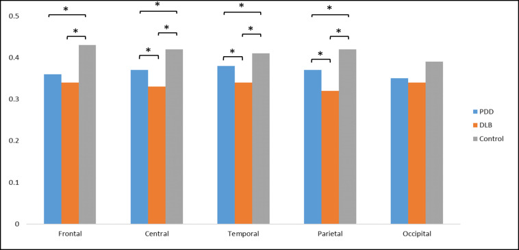
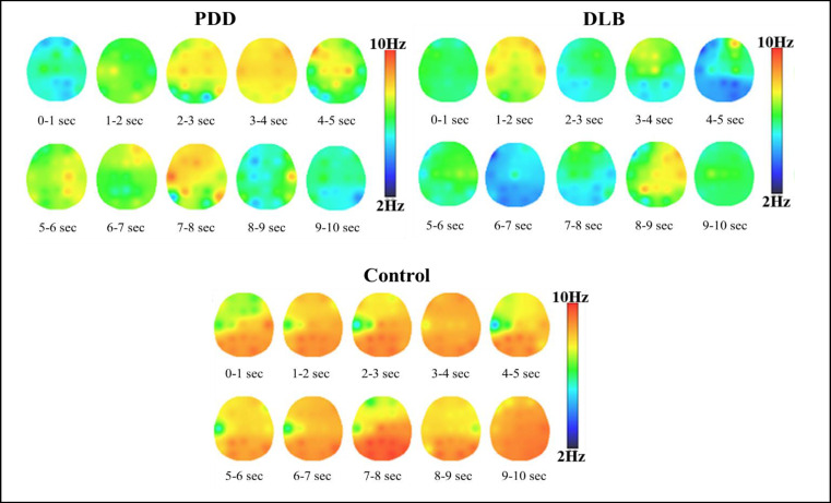
目的:帕金森病痴呆(PDD)和路易体痴呆(DLB)是突触核蛋白病分类的两种综合征,具有相似的症状。生物标记物的鉴定将为改善这些不同形式的痴呆的诊断、治疗和治疗效果监测提供一种准确的方法。方法:采用频谱分析和非线性动态分析比较PDD和DLB患者的脑电图特征。收集30例PDD患者、36例DLB患者和36例健康受试者静息时的脑电图数据。经过一个调节阶段以最小化噪声和消除伪像,我们使用Welch的方法和样本熵推导出光谱和复杂性特征。通过重复测量进行方差分析,比较两组之间大脑活动的频谱特征和非线性动态。结果:事后比较显示,与PDD和DLB患者相比,对照组的δ和θ波段功率较低,α和β波段功率较高。(p < 0.05)。在θ和α波段,PDD组的功率大于DLB组(P < 0.05)。此外,诊断的主效应显著(F = 4.67, P = 0.007),区域交互作用对复杂性值的诊断也显著(F = 4.58, P = 0.009)。事后分析显示,对照组在额区、中央区、颞区和顶叶区的脑电图复杂性显著高于PDD和DLB组(P < 0.05)。PDD组的中央、颞、顶叶脑电复杂度显著高于DLB组(P < 0.05)。结论:与对照组相比,PDD和DLB的脑电模式基本相似,但两者在脑电功率谱及其非线性动力学方面存在差异。我们的研究结果表明,与PDD相比,DLB患者在所有区域都有明显的弥漫性减缓和较低的皮层复杂性或活动,特别是在中央、颞叶和顶叶区域。
Comparison of Brain Activity between Patients with Parkinson Disease Dementia and Patients Affected by Dementia with Lewy Body through EEG Analysis.
Objective: Parkinson's disease dementia (PDD) and Dementia with Lewy bodies (DLB) are two syndromes categorized under synucleinopathy, sharing comparable symptoms. The identification of biomarkers would offer an accurate approach for improved diagnosis, treatment, and monitoring of treatment efficacy for these distinct forms of dementia. Method: This study utilized spectral analysis and nonlinear dynamic analysis to compare electroencephalogram (EEG) characteristics between PDD and DLB patients. EEG data was collected from 30 PDD patients, 36 DLB patients, and 36 healthy subjects at rest. Following a conditioning phase to minimize noise and eliminate artifacts, we derived spectral and complexity features using Welch's method and sample entropy. Analysis of variance with repeated measures was performed to compare spectral features and nonlinear dynamics of brain activity between the groups. Results: Post hoc comparison showed that in the control group, the power of delta and theta bands was lower and the power of alpha and beta bands was higher than in patients with PDD and DLB. (P < 0.05). In the theta and alpha bands, the PDD group showed greater power than the DLB group (P < 0.05). Furthermore, there was a significant main effect of diagnosis (F = 4.67, P = 0.007), and also the diagnosis by region interaction for complexity values (F = 4.58, P = 0.009). Post hoc analysis showed that the EEG complexity of the control group was significantly higher than that of the PDD and DLB groups in the frontal, central, temporal and parietal regions (P < 0.05). Moreover, the EEG complexity of the PDD group was significantly higher than that of the DLB group in the central, temporal and parietal regions (P < 0.05). Conclusion: Although both PDD and DLB had almost similar patterns compared to the control group, they showed differences in the EEG power spectrum and its nonlinear dynamics. Our findings indicated marked diffuse slowing and lower cortical complexity or activity in DLB patients compared to PDD in all regions, especially in the central, temporal and parietal areas.

 求助内容:
求助内容: 应助结果提醒方式:


