Silvia Bottazzi, Roberta V Ninkova, Luca Russo, Andrea Ponsiglione, Benedetta Gui, Daniela Demundo, Massimo Imbriaco, Aradhana M Venkatesan, Evis Sala, Stephanie Nougaret, Lucia Manganaro, Stefania Rizzo
{"title":"女性骨盆MRI检查妇科恶性肿瘤的偶然发现。","authors":"Silvia Bottazzi, Roberta V Ninkova, Luca Russo, Andrea Ponsiglione, Benedetta Gui, Daniela Demundo, Massimo Imbriaco, Aradhana M Venkatesan, Evis Sala, Stephanie Nougaret, Lucia Manganaro, Stefania Rizzo","doi":"10.1186/s13244-025-02006-5","DOIUrl":null,"url":null,"abstract":"<p><p>Incidental findings on female pelvic MRI present diagnostic challenges and may have significant clinical implications. Defined as abnormalities unrelated to the primary imaging indication, these findings have become increasingly prevalent with the expanded use of MRI in gynaecological practice. Standard gynaecological MRI protocols, incorporating T1- and T2-weighted sequences, diffusion-weighted imaging, and contrast-enhanced sequences, facilitate the characterisation of numerous extra-gynaecological abnormalities, ranging from benign to critical lesions. This review proposes a compartment-based approach for identifying extra-gynaecological findings, discussing their imaging characteristics and differential diagnoses. This approach may help radiologists systematically assess incidental findings, potentially improving the recognition of clinically relevant abnormalities and supporting timely clinical decision-making. CRITICAL RELEVANCE STATEMENT: Incidental extra-gynaecological findings on pelvic MRI can present significant diagnostic challenges. Systematic evaluation of incidental extra-gynaecological findings on pelvic MRI can improve radiologists' awareness of clinically relevant abnormalities. KEY POINTS: Extra-gynaecological incidental findings on pelvic MRI are common and range from benign to malignant conditions. A compartment-based classification-dividing the female pelvis into anterior, lateral, posterior, musculoskeletal, and miscellaneous compartments-provides a systematic framework for interpretation. Thorough assessment of all MRI sequences, including large field-of-view images, may help identify clinically relevant incidental findings.</p>","PeriodicalId":13639,"journal":{"name":"Insights into Imaging","volume":"16 1","pages":"143"},"PeriodicalIF":4.5000,"publicationDate":"2025-06-27","publicationTypes":"Journal Article","fieldsOfStudy":null,"isOpenAccess":false,"openAccessPdf":"https://www.ncbi.nlm.nih.gov/pmc/articles/PMC12204973/pdf/","citationCount":"0","resultStr":"{\"title\":\"Incidental findings in female pelvis MRI performed for gynaecological malignancies.\",\"authors\":\"Silvia Bottazzi, Roberta V Ninkova, Luca Russo, Andrea Ponsiglione, Benedetta Gui, Daniela Demundo, Massimo Imbriaco, Aradhana M Venkatesan, Evis Sala, Stephanie Nougaret, Lucia Manganaro, Stefania Rizzo\",\"doi\":\"10.1186/s13244-025-02006-5\",\"DOIUrl\":null,\"url\":null,\"abstract\":\"<p><p>Incidental findings on female pelvic MRI present diagnostic challenges and may have significant clinical implications. Defined as abnormalities unrelated to the primary imaging indication, these findings have become increasingly prevalent with the expanded use of MRI in gynaecological practice. Standard gynaecological MRI protocols, incorporating T1- and T2-weighted sequences, diffusion-weighted imaging, and contrast-enhanced sequences, facilitate the characterisation of numerous extra-gynaecological abnormalities, ranging from benign to critical lesions. This review proposes a compartment-based approach for identifying extra-gynaecological findings, discussing their imaging characteristics and differential diagnoses. This approach may help radiologists systematically assess incidental findings, potentially improving the recognition of clinically relevant abnormalities and supporting timely clinical decision-making. CRITICAL RELEVANCE STATEMENT: Incidental extra-gynaecological findings on pelvic MRI can present significant diagnostic challenges. Systematic evaluation of incidental extra-gynaecological findings on pelvic MRI can improve radiologists' awareness of clinically relevant abnormalities. KEY POINTS: Extra-gynaecological incidental findings on pelvic MRI are common and range from benign to malignant conditions. A compartment-based classification-dividing the female pelvis into anterior, lateral, posterior, musculoskeletal, and miscellaneous compartments-provides a systematic framework for interpretation. Thorough assessment of all MRI sequences, including large field-of-view images, may help identify clinically relevant incidental findings.</p>\",\"PeriodicalId\":13639,\"journal\":{\"name\":\"Insights into Imaging\",\"volume\":\"16 1\",\"pages\":\"143\"},\"PeriodicalIF\":4.5000,\"publicationDate\":\"2025-06-27\",\"publicationTypes\":\"Journal Article\",\"fieldsOfStudy\":null,\"isOpenAccess\":false,\"openAccessPdf\":\"https://www.ncbi.nlm.nih.gov/pmc/articles/PMC12204973/pdf/\",\"citationCount\":\"0\",\"resultStr\":null,\"platform\":\"Semanticscholar\",\"paperid\":null,\"PeriodicalName\":\"Insights into Imaging\",\"FirstCategoryId\":\"3\",\"ListUrlMain\":\"https://doi.org/10.1186/s13244-025-02006-5\",\"RegionNum\":2,\"RegionCategory\":\"医学\",\"ArticlePicture\":[],\"TitleCN\":null,\"AbstractTextCN\":null,\"PMCID\":null,\"EPubDate\":\"\",\"PubModel\":\"\",\"JCR\":\"Q1\",\"JCRName\":\"RADIOLOGY, NUCLEAR MEDICINE & MEDICAL IMAGING\",\"Score\":null,\"Total\":0}","platform":"Semanticscholar","paperid":null,"PeriodicalName":"Insights into Imaging","FirstCategoryId":"3","ListUrlMain":"https://doi.org/10.1186/s13244-025-02006-5","RegionNum":2,"RegionCategory":"医学","ArticlePicture":[],"TitleCN":null,"AbstractTextCN":null,"PMCID":null,"EPubDate":"","PubModel":"","JCR":"Q1","JCRName":"RADIOLOGY, NUCLEAR MEDICINE & MEDICAL IMAGING","Score":null,"Total":0}
Incidental findings in female pelvis MRI performed for gynaecological malignancies.
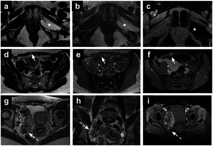
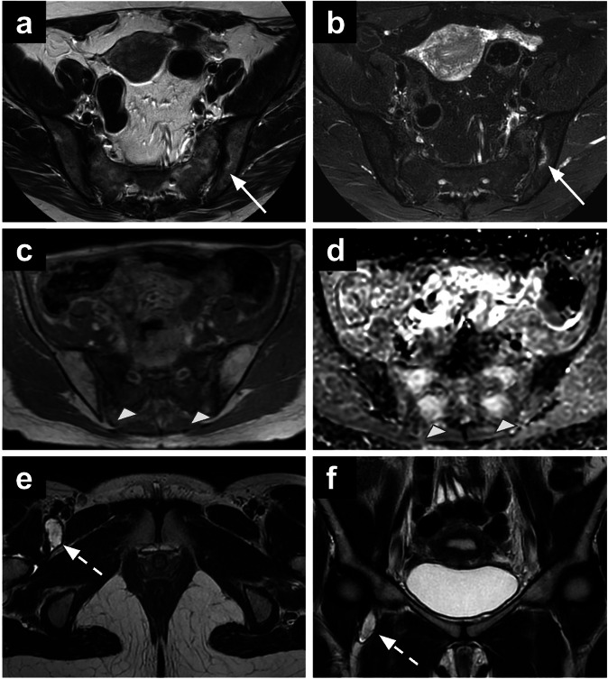
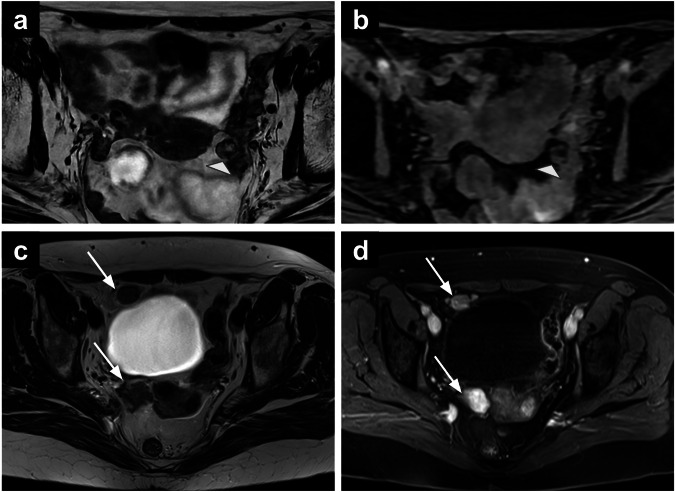
Incidental findings on female pelvic MRI present diagnostic challenges and may have significant clinical implications. Defined as abnormalities unrelated to the primary imaging indication, these findings have become increasingly prevalent with the expanded use of MRI in gynaecological practice. Standard gynaecological MRI protocols, incorporating T1- and T2-weighted sequences, diffusion-weighted imaging, and contrast-enhanced sequences, facilitate the characterisation of numerous extra-gynaecological abnormalities, ranging from benign to critical lesions. This review proposes a compartment-based approach for identifying extra-gynaecological findings, discussing their imaging characteristics and differential diagnoses. This approach may help radiologists systematically assess incidental findings, potentially improving the recognition of clinically relevant abnormalities and supporting timely clinical decision-making. CRITICAL RELEVANCE STATEMENT: Incidental extra-gynaecological findings on pelvic MRI can present significant diagnostic challenges. Systematic evaluation of incidental extra-gynaecological findings on pelvic MRI can improve radiologists' awareness of clinically relevant abnormalities. KEY POINTS: Extra-gynaecological incidental findings on pelvic MRI are common and range from benign to malignant conditions. A compartment-based classification-dividing the female pelvis into anterior, lateral, posterior, musculoskeletal, and miscellaneous compartments-provides a systematic framework for interpretation. Thorough assessment of all MRI sequences, including large field-of-view images, may help identify clinically relevant incidental findings.
期刊介绍:
Insights into Imaging (I³) is a peer-reviewed open access journal published under the brand SpringerOpen. All content published in the journal is freely available online to anyone, anywhere!
I³ continuously updates scientific knowledge and progress in best-practice standards in radiology through the publication of original articles and state-of-the-art reviews and opinions, along with recommendations and statements from the leading radiological societies in Europe.
Founded by the European Society of Radiology (ESR), I³ creates a platform for educational material, guidelines and recommendations, and a forum for topics of controversy.
A balanced combination of review articles, original papers, short communications from European radiological congresses and information on society matters makes I³ an indispensable source for current information in this field.
I³ is owned by the ESR, however authors retain copyright to their article according to the Creative Commons Attribution License (see Copyright and License Agreement). All articles can be read, redistributed and reused for free, as long as the author of the original work is cited properly.
The open access fees (article-processing charges) for this journal are kindly sponsored by ESR for all Members.
The journal went open access in 2012, which means that all articles published since then are freely available online.

 求助内容:
求助内容: 应助结果提醒方式:


