{"title":"可解释的多模态放射组学用于直肠癌肝转移的早期预测:一项多中心研究。","authors":"Yaru Feng, Jing Gong, Yanyan Wang, Yanfen Cui, Tong Tong","doi":"10.1186/s13244-025-02010-9","DOIUrl":null,"url":null,"abstract":"<p><strong>Objectives: </strong>To enhance liver metastasis (LM) risk prediction for rectal cancer (RC) patients using a multi-modal, explainable radiomics model based on rectal MRI and whole-liver CT, and to assess its prognostic value for survival.</p><p><strong>Methods: </strong>This retrospective study enrolled patients with pathologically confirmed RC from two medical centers. Radiomics features were extracted from rectal MRI as well as pre-metastatic liver CT. Feature selection was performed using ANOVA F-value and recursive feature elimination. The SHAP method elucidated the model's functionality by highlighting key feature contributions. Finally, Kaplan-Meier survival analysis and Cox regression assessed the prognostic utility of the model's prediction score.</p><p><strong>Results: </strong>A total of 431 patients were enrolled from two centers in our study. The radiomics model developed from baseline whole-liver CT features alone could predict LM development in all cohorts. A fusion model integrating liver CT with primary tumor MRI features provided synergetic effect and was more efficient in predicting LM, displaying an area under the receiver operating curve (AUC) of 0.85 (95% CI: 0.80-0.90) in the training cohort, and AUC values of 0.75 (95% CI: 0.64-0.86) and 0.73 (95% CI: 0.61-0.85) in the internal and external validation cohorts, respectively. SHAP summary plots illustrated how feature values influenced their impact on the model. The risk score generated by our model demonstrated significant prognostic value for LM-free survival (LMFS).</p><p><strong>Conclusions: </strong>The multi-modal, explainable radiomics model integrating primary tumor and pre-metastatic liver radiomics enhances the prediction of LM development and provides prognostic value in RC patients.</p><p><strong>Critical relevance statement: </strong>This study demonstrates that integrating radiomics features from pre-metastatic liver and primary tumors enhances the predictive performance for liver metastasis development in rectal cancer patients, highlighting its potential for personalized treatment planning and follow-up strategies for rectal cancer patients.</p><p><strong>Key points: </strong>Pre-metastatic liver CT radiomics features could predict the liver metastasis development of rectal cancer. Integrating primary tumor and pre-metastatic liver radiomics improved liver metastasis prediction accuracy. The model demonstrated favorable interpretability through SHAP method.</p>","PeriodicalId":13639,"journal":{"name":"Insights into Imaging","volume":"16 1","pages":"142"},"PeriodicalIF":4.5000,"publicationDate":"2025-06-27","publicationTypes":"Journal Article","fieldsOfStudy":null,"isOpenAccess":false,"openAccessPdf":"https://www.ncbi.nlm.nih.gov/pmc/articles/PMC12205110/pdf/","citationCount":"0","resultStr":"{\"title\":\"Explainable multi-modal radiomics for early prediction of liver metastasis in rectal cancer: a multicentric study.\",\"authors\":\"Yaru Feng, Jing Gong, Yanyan Wang, Yanfen Cui, Tong Tong\",\"doi\":\"10.1186/s13244-025-02010-9\",\"DOIUrl\":null,\"url\":null,\"abstract\":\"<p><strong>Objectives: </strong>To enhance liver metastasis (LM) risk prediction for rectal cancer (RC) patients using a multi-modal, explainable radiomics model based on rectal MRI and whole-liver CT, and to assess its prognostic value for survival.</p><p><strong>Methods: </strong>This retrospective study enrolled patients with pathologically confirmed RC from two medical centers. Radiomics features were extracted from rectal MRI as well as pre-metastatic liver CT. Feature selection was performed using ANOVA F-value and recursive feature elimination. The SHAP method elucidated the model's functionality by highlighting key feature contributions. Finally, Kaplan-Meier survival analysis and Cox regression assessed the prognostic utility of the model's prediction score.</p><p><strong>Results: </strong>A total of 431 patients were enrolled from two centers in our study. The radiomics model developed from baseline whole-liver CT features alone could predict LM development in all cohorts. A fusion model integrating liver CT with primary tumor MRI features provided synergetic effect and was more efficient in predicting LM, displaying an area under the receiver operating curve (AUC) of 0.85 (95% CI: 0.80-0.90) in the training cohort, and AUC values of 0.75 (95% CI: 0.64-0.86) and 0.73 (95% CI: 0.61-0.85) in the internal and external validation cohorts, respectively. SHAP summary plots illustrated how feature values influenced their impact on the model. The risk score generated by our model demonstrated significant prognostic value for LM-free survival (LMFS).</p><p><strong>Conclusions: </strong>The multi-modal, explainable radiomics model integrating primary tumor and pre-metastatic liver radiomics enhances the prediction of LM development and provides prognostic value in RC patients.</p><p><strong>Critical relevance statement: </strong>This study demonstrates that integrating radiomics features from pre-metastatic liver and primary tumors enhances the predictive performance for liver metastasis development in rectal cancer patients, highlighting its potential for personalized treatment planning and follow-up strategies for rectal cancer patients.</p><p><strong>Key points: </strong>Pre-metastatic liver CT radiomics features could predict the liver metastasis development of rectal cancer. Integrating primary tumor and pre-metastatic liver radiomics improved liver metastasis prediction accuracy. The model demonstrated favorable interpretability through SHAP method.</p>\",\"PeriodicalId\":13639,\"journal\":{\"name\":\"Insights into Imaging\",\"volume\":\"16 1\",\"pages\":\"142\"},\"PeriodicalIF\":4.5000,\"publicationDate\":\"2025-06-27\",\"publicationTypes\":\"Journal Article\",\"fieldsOfStudy\":null,\"isOpenAccess\":false,\"openAccessPdf\":\"https://www.ncbi.nlm.nih.gov/pmc/articles/PMC12205110/pdf/\",\"citationCount\":\"0\",\"resultStr\":null,\"platform\":\"Semanticscholar\",\"paperid\":null,\"PeriodicalName\":\"Insights into Imaging\",\"FirstCategoryId\":\"3\",\"ListUrlMain\":\"https://doi.org/10.1186/s13244-025-02010-9\",\"RegionNum\":2,\"RegionCategory\":\"医学\",\"ArticlePicture\":[],\"TitleCN\":null,\"AbstractTextCN\":null,\"PMCID\":null,\"EPubDate\":\"\",\"PubModel\":\"\",\"JCR\":\"Q1\",\"JCRName\":\"RADIOLOGY, NUCLEAR MEDICINE & MEDICAL IMAGING\",\"Score\":null,\"Total\":0}","platform":"Semanticscholar","paperid":null,"PeriodicalName":"Insights into Imaging","FirstCategoryId":"3","ListUrlMain":"https://doi.org/10.1186/s13244-025-02010-9","RegionNum":2,"RegionCategory":"医学","ArticlePicture":[],"TitleCN":null,"AbstractTextCN":null,"PMCID":null,"EPubDate":"","PubModel":"","JCR":"Q1","JCRName":"RADIOLOGY, NUCLEAR MEDICINE & MEDICAL IMAGING","Score":null,"Total":0}
Explainable multi-modal radiomics for early prediction of liver metastasis in rectal cancer: a multicentric study.
Objectives: To enhance liver metastasis (LM) risk prediction for rectal cancer (RC) patients using a multi-modal, explainable radiomics model based on rectal MRI and whole-liver CT, and to assess its prognostic value for survival.
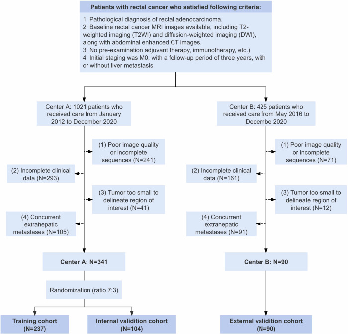
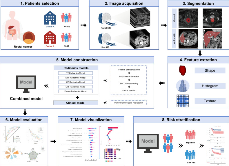
Methods: This retrospective study enrolled patients with pathologically confirmed RC from two medical centers. Radiomics features were extracted from rectal MRI as well as pre-metastatic liver CT. Feature selection was performed using ANOVA F-value and recursive feature elimination. The SHAP method elucidated the model's functionality by highlighting key feature contributions. Finally, Kaplan-Meier survival analysis and Cox regression assessed the prognostic utility of the model's prediction score.
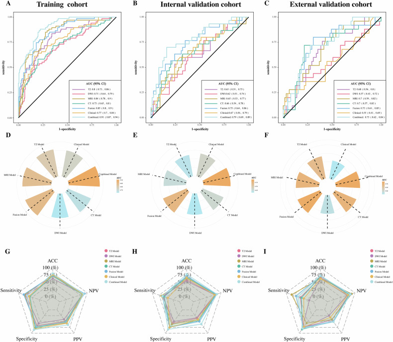
Results: A total of 431 patients were enrolled from two centers in our study. The radiomics model developed from baseline whole-liver CT features alone could predict LM development in all cohorts. A fusion model integrating liver CT with primary tumor MRI features provided synergetic effect and was more efficient in predicting LM, displaying an area under the receiver operating curve (AUC) of 0.85 (95% CI: 0.80-0.90) in the training cohort, and AUC values of 0.75 (95% CI: 0.64-0.86) and 0.73 (95% CI: 0.61-0.85) in the internal and external validation cohorts, respectively. SHAP summary plots illustrated how feature values influenced their impact on the model. The risk score generated by our model demonstrated significant prognostic value for LM-free survival (LMFS).
Conclusions: The multi-modal, explainable radiomics model integrating primary tumor and pre-metastatic liver radiomics enhances the prediction of LM development and provides prognostic value in RC patients.
Critical relevance statement: This study demonstrates that integrating radiomics features from pre-metastatic liver and primary tumors enhances the predictive performance for liver metastasis development in rectal cancer patients, highlighting its potential for personalized treatment planning and follow-up strategies for rectal cancer patients.
Key points: Pre-metastatic liver CT radiomics features could predict the liver metastasis development of rectal cancer. Integrating primary tumor and pre-metastatic liver radiomics improved liver metastasis prediction accuracy. The model demonstrated favorable interpretability through SHAP method.
期刊介绍:
Insights into Imaging (I³) is a peer-reviewed open access journal published under the brand SpringerOpen. All content published in the journal is freely available online to anyone, anywhere!
I³ continuously updates scientific knowledge and progress in best-practice standards in radiology through the publication of original articles and state-of-the-art reviews and opinions, along with recommendations and statements from the leading radiological societies in Europe.
Founded by the European Society of Radiology (ESR), I³ creates a platform for educational material, guidelines and recommendations, and a forum for topics of controversy.
A balanced combination of review articles, original papers, short communications from European radiological congresses and information on society matters makes I³ an indispensable source for current information in this field.
I³ is owned by the ESR, however authors retain copyright to their article according to the Creative Commons Attribution License (see Copyright and License Agreement). All articles can be read, redistributed and reused for free, as long as the author of the original work is cited properly.
The open access fees (article-processing charges) for this journal are kindly sponsored by ESR for all Members.
The journal went open access in 2012, which means that all articles published since then are freely available online.

 求助内容:
求助内容: 应助结果提醒方式:


