Basma Baccouche, Maximilian McCann, Janani Rajasekar, Nguyễn Thị Thanh Nhàn, Kaori Yamada, Andrius Kazlauskas
{"title":"激活素A通过干扰活化的VEGFR2的运输来防止病理血管中对VEGF的高反应性。","authors":"Basma Baccouche, Maximilian McCann, Janani Rajasekar, Nguyễn Thị Thanh Nhàn, Kaori Yamada, Andrius Kazlauskas","doi":"10.1016/j.ajpath.2025.05.022","DOIUrl":null,"url":null,"abstract":"<p><p>Although quality-of-life compromising afflictions, such as diabetic retinopathy (DR), are driven by vascular endothelial growth factor (VEGF), there is a growing appreciation that the concentration of VEGF is not the only influencer of vascular dysfunction within the retina. Activin A (activin), a ligand of the transforming growth factor-β superfamily, attenuated VEGF-induced vascular endothelial-cadherin disorganization, pore formation, and permeability of primary human retinal endothelial cells. Efforts to further investigate the mechanism of this phenomenon revealed that activin reduced the expression of Rab11, which was required for the activin effect. The activin effect was not observed in cells with suppressed expression of the endosomally localized protein tyrosine phosphatase (PTP1b), whereas protein tyrosine phosphatases present on the plasma membrane were dispensable. Activin attenuated VEGF-mediated phosphorylation of VEGF receptor 2 at 30 minutes and longer post-stimulation time points. Together, these data support the concept that activin suppressed VEGF-induced barrier relaxation by perturbing trafficking of activated VEGF receptor 2. This activin-based suppression of responsiveness to VEGF was compromised in the endothelium of pathologic blood vessels from patients who developed end-stage proliferative DR. This defect rendered human retinal endothelial cell hyperresponsive to VEGF and was not observed in retinal vessels of diabetic mice, which do not develop the angiogenic forms of DR. These data provide novel insights regarding the pathogenesis of proliferative DR in patients.</p>","PeriodicalId":7623,"journal":{"name":"American Journal of Pathology","volume":" ","pages":""},"PeriodicalIF":4.7000,"publicationDate":"2025-06-24","publicationTypes":"Journal Article","fieldsOfStudy":null,"isOpenAccess":false,"openAccessPdf":"","citationCount":"0","resultStr":"{\"title\":\"Activin A Prevents Hyperresponsiveness to Vascular Endothelial Growth Factor in Pathologic Blood Vessels by Perturbing the Trafficking of Activated Vascular Endothelial Growth Factor Receptor 2.\",\"authors\":\"Basma Baccouche, Maximilian McCann, Janani Rajasekar, Nguyễn Thị Thanh Nhàn, Kaori Yamada, Andrius Kazlauskas\",\"doi\":\"10.1016/j.ajpath.2025.05.022\",\"DOIUrl\":null,\"url\":null,\"abstract\":\"<p><p>Although quality-of-life compromising afflictions, such as diabetic retinopathy (DR), are driven by vascular endothelial growth factor (VEGF), there is a growing appreciation that the concentration of VEGF is not the only influencer of vascular dysfunction within the retina. Activin A (activin), a ligand of the transforming growth factor-β superfamily, attenuated VEGF-induced vascular endothelial-cadherin disorganization, pore formation, and permeability of primary human retinal endothelial cells. Efforts to further investigate the mechanism of this phenomenon revealed that activin reduced the expression of Rab11, which was required for the activin effect. The activin effect was not observed in cells with suppressed expression of the endosomally localized protein tyrosine phosphatase (PTP1b), whereas protein tyrosine phosphatases present on the plasma membrane were dispensable. Activin attenuated VEGF-mediated phosphorylation of VEGF receptor 2 at 30 minutes and longer post-stimulation time points. Together, these data support the concept that activin suppressed VEGF-induced barrier relaxation by perturbing trafficking of activated VEGF receptor 2. This activin-based suppression of responsiveness to VEGF was compromised in the endothelium of pathologic blood vessels from patients who developed end-stage proliferative DR. This defect rendered human retinal endothelial cell hyperresponsive to VEGF and was not observed in retinal vessels of diabetic mice, which do not develop the angiogenic forms of DR. These data provide novel insights regarding the pathogenesis of proliferative DR in patients.</p>\",\"PeriodicalId\":7623,\"journal\":{\"name\":\"American Journal of Pathology\",\"volume\":\" \",\"pages\":\"\"},\"PeriodicalIF\":4.7000,\"publicationDate\":\"2025-06-24\",\"publicationTypes\":\"Journal Article\",\"fieldsOfStudy\":null,\"isOpenAccess\":false,\"openAccessPdf\":\"\",\"citationCount\":\"0\",\"resultStr\":null,\"platform\":\"Semanticscholar\",\"paperid\":null,\"PeriodicalName\":\"American Journal of Pathology\",\"FirstCategoryId\":\"3\",\"ListUrlMain\":\"https://doi.org/10.1016/j.ajpath.2025.05.022\",\"RegionNum\":2,\"RegionCategory\":\"医学\",\"ArticlePicture\":[],\"TitleCN\":null,\"AbstractTextCN\":null,\"PMCID\":null,\"EPubDate\":\"\",\"PubModel\":\"\",\"JCR\":\"Q1\",\"JCRName\":\"PATHOLOGY\",\"Score\":null,\"Total\":0}","platform":"Semanticscholar","paperid":null,"PeriodicalName":"American Journal of Pathology","FirstCategoryId":"3","ListUrlMain":"https://doi.org/10.1016/j.ajpath.2025.05.022","RegionNum":2,"RegionCategory":"医学","ArticlePicture":[],"TitleCN":null,"AbstractTextCN":null,"PMCID":null,"EPubDate":"","PubModel":"","JCR":"Q1","JCRName":"PATHOLOGY","Score":null,"Total":0}
Activin A Prevents Hyperresponsiveness to Vascular Endothelial Growth Factor in Pathologic Blood Vessels by Perturbing the Trafficking of Activated Vascular Endothelial Growth Factor Receptor 2.
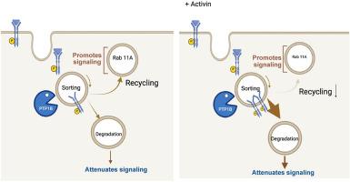
Although quality-of-life compromising afflictions, such as diabetic retinopathy (DR), are driven by vascular endothelial growth factor (VEGF), there is a growing appreciation that the concentration of VEGF is not the only influencer of vascular dysfunction within the retina. Activin A (activin), a ligand of the transforming growth factor-β superfamily, attenuated VEGF-induced vascular endothelial-cadherin disorganization, pore formation, and permeability of primary human retinal endothelial cells. Efforts to further investigate the mechanism of this phenomenon revealed that activin reduced the expression of Rab11, which was required for the activin effect. The activin effect was not observed in cells with suppressed expression of the endosomally localized protein tyrosine phosphatase (PTP1b), whereas protein tyrosine phosphatases present on the plasma membrane were dispensable. Activin attenuated VEGF-mediated phosphorylation of VEGF receptor 2 at 30 minutes and longer post-stimulation time points. Together, these data support the concept that activin suppressed VEGF-induced barrier relaxation by perturbing trafficking of activated VEGF receptor 2. This activin-based suppression of responsiveness to VEGF was compromised in the endothelium of pathologic blood vessels from patients who developed end-stage proliferative DR. This defect rendered human retinal endothelial cell hyperresponsive to VEGF and was not observed in retinal vessels of diabetic mice, which do not develop the angiogenic forms of DR. These data provide novel insights regarding the pathogenesis of proliferative DR in patients.
期刊介绍:
The American Journal of Pathology, official journal of the American Society for Investigative Pathology, published by Elsevier, Inc., seeks high-quality original research reports, reviews, and commentaries related to the molecular and cellular basis of disease. The editors will consider basic, translational, and clinical investigations that directly address mechanisms of pathogenesis or provide a foundation for future mechanistic inquiries. Examples of such foundational investigations include data mining, identification of biomarkers, molecular pathology, and discovery research. Foundational studies that incorporate deep learning and artificial intelligence are also welcome. High priority is given to studies of human disease and relevant experimental models using molecular, cellular, and organismal approaches.