Balazs C Lengyel, Ponraj Chinnadurai, Rebecca G Barnes, Charudatta S Bavare, Alan B Lumsden
{"title":"CT图像引导的血管手术机器人的早期概念:在尸体模型模拟过程中腹膜后结构的移位。","authors":"Balazs C Lengyel, Ponraj Chinnadurai, Rebecca G Barnes, Charudatta S Bavare, Alan B Lumsden","doi":"10.3390/tomography11060060","DOIUrl":null,"url":null,"abstract":"<p><strong>Background: </strong>CT image guidance and navigation, although routinely used in complex endovascular procedures, is an unexplored territory in evolving vascular robotic procedures. In robotic surgery, it promises the better localization of vasculature, the optimization of port placement, less inadvertent tissue damage, and increased patient safety during the dissection of retroperitoneal structures. However, unknown tissue displacement resulting from induced pneumoperitoneum and positional changes compared to the preoperative CT scan can pose significant limitations to the reliability of image guidance. We aimed to study the displacement of retroperitoneal organs and vasculature due to factors such as increased intra-abdominal pressure (IAP) due to CO<sub>2</sub> insufflation and patient positioning (PP) using intraoperative CT imaging in a cadaveric model.</p><p><strong>Methods: </strong>A thawed, fresh-frozen human cadaveric model was positioned according to simulated procedural workflows. Intra-arterial, contrast-enhanced CT scans were performed after the insertion of four laparoscopic ports in the abdomen. CT scans were performed with 0-5-15-25 mmHg IAPs in supine, left lateral decubitus, right lateral decubitus, Trendelenburg, and reverse Trendelenburg positions. Euclidean distances between fixed anatomical bony and retroperitoneal vascular landmarks were measured and compared across different CT scans.</p><p><strong>Results: </strong>Comparing the effects of various IAPs to the baseline (zero IAP) in the same PP, an average displacement for retroperitoneal vascular landmarks ranged from 0.6 to 3.0 mm (SD 1.0-2.8 mm). When changing the PPs while maintaining the same IAP, the average displacement of the retroperitoneal vasculature ranged from 2.0 to 15.0 mm (SD 1.7-7.2 mm).</p><p><strong>Conclusions: </strong>Our preliminary imaging findings from a single cadaveric model suggest minimal (~3 mm maximum) target vasculature displacement in the retroperitoneum due to elevated IAP in supine position and higher displacement due to changes in patient positioning. Similar imaging studies are needed to quantify procedural workflow-specific and anatomy-specific deformation, which would be invaluable in developing and validating advanced tissue deformation models, facilitating the routine applicability and usefulness of CT image guidance for target delineation during robotic vascular procedures.</p>","PeriodicalId":51330,"journal":{"name":"Tomography","volume":"11 6","pages":""},"PeriodicalIF":2.2000,"publicationDate":"2025-05-23","publicationTypes":"Journal Article","fieldsOfStudy":null,"isOpenAccess":false,"openAccessPdf":"https://www.ncbi.nlm.nih.gov/pmc/articles/PMC12197135/pdf/","citationCount":"0","resultStr":"{\"title\":\"Early Concepts in CT Image-Guided Robotic Vascular Surgery: The Displacement of Retroperitoneal Structures During Simulated Procedures in a Cadaveric Model.\",\"authors\":\"Balazs C Lengyel, Ponraj Chinnadurai, Rebecca G Barnes, Charudatta S Bavare, Alan B Lumsden\",\"doi\":\"10.3390/tomography11060060\",\"DOIUrl\":null,\"url\":null,\"abstract\":\"<p><strong>Background: </strong>CT image guidance and navigation, although routinely used in complex endovascular procedures, is an unexplored territory in evolving vascular robotic procedures. In robotic surgery, it promises the better localization of vasculature, the optimization of port placement, less inadvertent tissue damage, and increased patient safety during the dissection of retroperitoneal structures. However, unknown tissue displacement resulting from induced pneumoperitoneum and positional changes compared to the preoperative CT scan can pose significant limitations to the reliability of image guidance. We aimed to study the displacement of retroperitoneal organs and vasculature due to factors such as increased intra-abdominal pressure (IAP) due to CO<sub>2</sub> insufflation and patient positioning (PP) using intraoperative CT imaging in a cadaveric model.</p><p><strong>Methods: </strong>A thawed, fresh-frozen human cadaveric model was positioned according to simulated procedural workflows. Intra-arterial, contrast-enhanced CT scans were performed after the insertion of four laparoscopic ports in the abdomen. CT scans were performed with 0-5-15-25 mmHg IAPs in supine, left lateral decubitus, right lateral decubitus, Trendelenburg, and reverse Trendelenburg positions. Euclidean distances between fixed anatomical bony and retroperitoneal vascular landmarks were measured and compared across different CT scans.</p><p><strong>Results: </strong>Comparing the effects of various IAPs to the baseline (zero IAP) in the same PP, an average displacement for retroperitoneal vascular landmarks ranged from 0.6 to 3.0 mm (SD 1.0-2.8 mm). When changing the PPs while maintaining the same IAP, the average displacement of the retroperitoneal vasculature ranged from 2.0 to 15.0 mm (SD 1.7-7.2 mm).</p><p><strong>Conclusions: </strong>Our preliminary imaging findings from a single cadaveric model suggest minimal (~3 mm maximum) target vasculature displacement in the retroperitoneum due to elevated IAP in supine position and higher displacement due to changes in patient positioning. Similar imaging studies are needed to quantify procedural workflow-specific and anatomy-specific deformation, which would be invaluable in developing and validating advanced tissue deformation models, facilitating the routine applicability and usefulness of CT image guidance for target delineation during robotic vascular procedures.</p>\",\"PeriodicalId\":51330,\"journal\":{\"name\":\"Tomography\",\"volume\":\"11 6\",\"pages\":\"\"},\"PeriodicalIF\":2.2000,\"publicationDate\":\"2025-05-23\",\"publicationTypes\":\"Journal Article\",\"fieldsOfStudy\":null,\"isOpenAccess\":false,\"openAccessPdf\":\"https://www.ncbi.nlm.nih.gov/pmc/articles/PMC12197135/pdf/\",\"citationCount\":\"0\",\"resultStr\":null,\"platform\":\"Semanticscholar\",\"paperid\":null,\"PeriodicalName\":\"Tomography\",\"FirstCategoryId\":\"3\",\"ListUrlMain\":\"https://doi.org/10.3390/tomography11060060\",\"RegionNum\":4,\"RegionCategory\":\"医学\",\"ArticlePicture\":[],\"TitleCN\":null,\"AbstractTextCN\":null,\"PMCID\":null,\"EPubDate\":\"\",\"PubModel\":\"\",\"JCR\":\"Q2\",\"JCRName\":\"RADIOLOGY, NUCLEAR MEDICINE & MEDICAL IMAGING\",\"Score\":null,\"Total\":0}","platform":"Semanticscholar","paperid":null,"PeriodicalName":"Tomography","FirstCategoryId":"3","ListUrlMain":"https://doi.org/10.3390/tomography11060060","RegionNum":4,"RegionCategory":"医学","ArticlePicture":[],"TitleCN":null,"AbstractTextCN":null,"PMCID":null,"EPubDate":"","PubModel":"","JCR":"Q2","JCRName":"RADIOLOGY, NUCLEAR MEDICINE & MEDICAL IMAGING","Score":null,"Total":0}
引用次数: 0
摘要
背景:CT图像引导和导航虽然经常用于复杂的血管内手术,但在不断发展的血管机器人手术中仍是一个未开发的领域。在机器人手术中,它有望更好地定位血管系统,优化端口放置,减少无意的组织损伤,并增加患者在腹膜后结构解剖过程中的安全性。然而,与术前CT扫描相比,由诱导气腹和位置变化引起的未知组织移位会对图像引导的可靠性造成重大限制。我们的目的是研究腹膜后器官和脉管系统的位移,由于诸如增加腹内压(IAP)由于二氧化碳的注入和患者的体位(PP)在尸体模型中使用术中CT成像。方法:根据模拟的程序工作流程定位解冻、新鲜冷冻的人体尸体模型。在腹部插入四个腹腔镜端口后进行动脉内对比增强CT扫描。采用0-5-15-25 mmHg IAPs在仰卧位、左侧侧卧位、右侧侧卧位、Trendelenburg位和反向Trendelenburg位进行CT扫描。在不同的CT扫描中测量和比较固定解剖骨和腹膜后血管标志之间的欧几里得距离。结果:在同一PP中,将不同IAP与基线(零IAP)的效果进行比较,腹膜后血管地标的平均位移范围为0.6至3.0 mm (SD 1.0-2.8 mm)。在保持IAP不变的情况下改变PPs,腹膜后血管的平均位移为2.0 ~ 15.0 mm (SD 1.7 ~ 7.2 mm)。结论:我们对单个尸体模型的初步成像结果显示,由于仰卧位时IAP升高,腹膜后靶血管位移最小(最大约3mm),而由于患者体位的改变,靶血管位移更大。需要类似的成像研究来量化程序工作流程特定和解剖结构特定的变形,这对于开发和验证高级组织变形模型将是非常宝贵的,有助于在机器人血管手术过程中CT图像指导目标描绘的常规适用性和实用性。
Early Concepts in CT Image-Guided Robotic Vascular Surgery: The Displacement of Retroperitoneal Structures During Simulated Procedures in a Cadaveric Model.
Background: CT image guidance and navigation, although routinely used in complex endovascular procedures, is an unexplored territory in evolving vascular robotic procedures. In robotic surgery, it promises the better localization of vasculature, the optimization of port placement, less inadvertent tissue damage, and increased patient safety during the dissection of retroperitoneal structures. However, unknown tissue displacement resulting from induced pneumoperitoneum and positional changes compared to the preoperative CT scan can pose significant limitations to the reliability of image guidance. We aimed to study the displacement of retroperitoneal organs and vasculature due to factors such as increased intra-abdominal pressure (IAP) due to CO2 insufflation and patient positioning (PP) using intraoperative CT imaging in a cadaveric model.
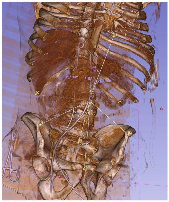
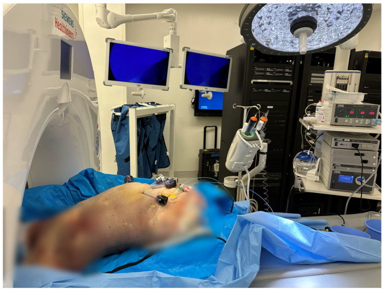
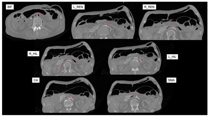
Methods: A thawed, fresh-frozen human cadaveric model was positioned according to simulated procedural workflows. Intra-arterial, contrast-enhanced CT scans were performed after the insertion of four laparoscopic ports in the abdomen. CT scans were performed with 0-5-15-25 mmHg IAPs in supine, left lateral decubitus, right lateral decubitus, Trendelenburg, and reverse Trendelenburg positions. Euclidean distances between fixed anatomical bony and retroperitoneal vascular landmarks were measured and compared across different CT scans.
Results: Comparing the effects of various IAPs to the baseline (zero IAP) in the same PP, an average displacement for retroperitoneal vascular landmarks ranged from 0.6 to 3.0 mm (SD 1.0-2.8 mm). When changing the PPs while maintaining the same IAP, the average displacement of the retroperitoneal vasculature ranged from 2.0 to 15.0 mm (SD 1.7-7.2 mm).
Conclusions: Our preliminary imaging findings from a single cadaveric model suggest minimal (~3 mm maximum) target vasculature displacement in the retroperitoneum due to elevated IAP in supine position and higher displacement due to changes in patient positioning. Similar imaging studies are needed to quantify procedural workflow-specific and anatomy-specific deformation, which would be invaluable in developing and validating advanced tissue deformation models, facilitating the routine applicability and usefulness of CT image guidance for target delineation during robotic vascular procedures.
TomographyMedicine-Radiology, Nuclear Medicine and Imaging
CiteScore
2.70
自引率
10.50%
发文量
222
期刊介绍:
TomographyTM publishes basic (technical and pre-clinical) and clinical scientific articles which involve the advancement of imaging technologies. Tomography encompasses studies that use single or multiple imaging modalities including for example CT, US, PET, SPECT, MR and hyperpolarization technologies, as well as optical modalities (i.e. bioluminescence, photoacoustic, endomicroscopy, fiber optic imaging and optical computed tomography) in basic sciences, engineering, preclinical and clinical medicine.
Tomography also welcomes studies involving exploration and refinement of contrast mechanisms and image-derived metrics within and across modalities toward the development of novel imaging probes for image-based feedback and intervention. The use of imaging in biology and medicine provides unparalleled opportunities to noninvasively interrogate tissues to obtain real-time dynamic and quantitative information required for diagnosis and response to interventions and to follow evolving pathological conditions. As multi-modal studies and the complexities of imaging technologies themselves are ever increasing to provide advanced information to scientists and clinicians.
Tomography provides a unique publication venue allowing investigators the opportunity to more precisely communicate integrated findings related to the diverse and heterogeneous features associated with underlying anatomical, physiological, functional, metabolic and molecular genetic activities of normal and diseased tissue. Thus Tomography publishes peer-reviewed articles which involve the broad use of imaging of any tissue and disease type including both preclinical and clinical investigations. In addition, hardware/software along with chemical and molecular probe advances are welcome as they are deemed to significantly contribute towards the long-term goal of improving the overall impact of imaging on scientific and clinical discovery.

 求助内容:
求助内容: 应助结果提醒方式:


