Joohyun Lee, Jee Young Lee, Keum Nae Kang, Jae Ni Jang, Sukhee Park, Young Uk Kim
{"title":"确定C5/6水平椎间孔横截面积最合适的截断点以预测颈椎椎间孔骨狭窄。","authors":"Joohyun Lee, Jee Young Lee, Keum Nae Kang, Jae Ni Jang, Sukhee Park, Young Uk Kim","doi":"10.3390/tomography11060067","DOIUrl":null,"url":null,"abstract":"<p><p><b>Background</b>: Cervical foraminal bony stenosis (CFBS) is a common degenerative spinal condition that causes radicular pain and functional impairment in the upper extremities. Accurate and objective diagnosis of CFBS remains challenging due to the absence of standardized morphometric criteria. This study aimed to determine an optimal cut-off value for the cervical foraminal cross-sectional area (CFCSA) at the C5/6 level as a diagnostic indicator of CFBS. <b>Methods</b>: We conducted a retrospective case-control study including 154 patients aged 50 years or older with clinically and radiologically confirmed CFBS and 150 age-matched asymptomatic controls. Cervical spine magnetic resonance imaging (MRI) was performed in all subjects and CFCSA measurements were obtained from sagittal T2-weighted images using a standardized protocol. Group differences were analyzed using <i>t</i>-tests and diagnostic performance was assessed using receiver operating characteristic (ROC) curve analysis. <b>Results</b>: The mean CFCSA was significantly lower in the CFBS group (25.65 ± 7.19 mm<sup>2</sup>) compared to the control group (43.00 ± 8.38 mm<sup>2</sup>; <i>p</i> < 0.001). ROC analysis identified a CFCSA threshold of 33.02 mm<sup>2</sup> as the optimal cut-off point for predicting CFBS, yielding a sensitivity of 86.4%, a specificity of 86.7%, and an area under the curve (AUC) of 0.94 (95% CI: 0.91-0.96). <b>Conclusions</b>: These findings suggest that CFCSA is a robust and reproducible morphological parameter for evaluating foraminal stenosis. The proposed cut-off may enhance diagnostic accuracy and aid in clinical decision-making for patients presenting with C6 radiculopathy. However, given this study's retrospective, single-center design, further validation through multicenter, prospective studies across multiple cervical levels is warranted.</p>","PeriodicalId":51330,"journal":{"name":"Tomography","volume":"11 6","pages":""},"PeriodicalIF":2.2000,"publicationDate":"2025-06-10","publicationTypes":"Journal Article","fieldsOfStudy":null,"isOpenAccess":false,"openAccessPdf":"https://www.ncbi.nlm.nih.gov/pmc/articles/PMC12196652/pdf/","citationCount":"0","resultStr":"{\"title\":\"Determination of the Most Suitable Cut-Off Point of the Cervical Foraminal Cross-Sectional Area at the C5/6 Level to Predict Cervical Foraminal Bony Stenosis.\",\"authors\":\"Joohyun Lee, Jee Young Lee, Keum Nae Kang, Jae Ni Jang, Sukhee Park, Young Uk Kim\",\"doi\":\"10.3390/tomography11060067\",\"DOIUrl\":null,\"url\":null,\"abstract\":\"<p><p><b>Background</b>: Cervical foraminal bony stenosis (CFBS) is a common degenerative spinal condition that causes radicular pain and functional impairment in the upper extremities. Accurate and objective diagnosis of CFBS remains challenging due to the absence of standardized morphometric criteria. This study aimed to determine an optimal cut-off value for the cervical foraminal cross-sectional area (CFCSA) at the C5/6 level as a diagnostic indicator of CFBS. <b>Methods</b>: We conducted a retrospective case-control study including 154 patients aged 50 years or older with clinically and radiologically confirmed CFBS and 150 age-matched asymptomatic controls. Cervical spine magnetic resonance imaging (MRI) was performed in all subjects and CFCSA measurements were obtained from sagittal T2-weighted images using a standardized protocol. Group differences were analyzed using <i>t</i>-tests and diagnostic performance was assessed using receiver operating characteristic (ROC) curve analysis. <b>Results</b>: The mean CFCSA was significantly lower in the CFBS group (25.65 ± 7.19 mm<sup>2</sup>) compared to the control group (43.00 ± 8.38 mm<sup>2</sup>; <i>p</i> < 0.001). ROC analysis identified a CFCSA threshold of 33.02 mm<sup>2</sup> as the optimal cut-off point for predicting CFBS, yielding a sensitivity of 86.4%, a specificity of 86.7%, and an area under the curve (AUC) of 0.94 (95% CI: 0.91-0.96). <b>Conclusions</b>: These findings suggest that CFCSA is a robust and reproducible morphological parameter for evaluating foraminal stenosis. The proposed cut-off may enhance diagnostic accuracy and aid in clinical decision-making for patients presenting with C6 radiculopathy. However, given this study's retrospective, single-center design, further validation through multicenter, prospective studies across multiple cervical levels is warranted.</p>\",\"PeriodicalId\":51330,\"journal\":{\"name\":\"Tomography\",\"volume\":\"11 6\",\"pages\":\"\"},\"PeriodicalIF\":2.2000,\"publicationDate\":\"2025-06-10\",\"publicationTypes\":\"Journal Article\",\"fieldsOfStudy\":null,\"isOpenAccess\":false,\"openAccessPdf\":\"https://www.ncbi.nlm.nih.gov/pmc/articles/PMC12196652/pdf/\",\"citationCount\":\"0\",\"resultStr\":null,\"platform\":\"Semanticscholar\",\"paperid\":null,\"PeriodicalName\":\"Tomography\",\"FirstCategoryId\":\"3\",\"ListUrlMain\":\"https://doi.org/10.3390/tomography11060067\",\"RegionNum\":4,\"RegionCategory\":\"医学\",\"ArticlePicture\":[],\"TitleCN\":null,\"AbstractTextCN\":null,\"PMCID\":null,\"EPubDate\":\"\",\"PubModel\":\"\",\"JCR\":\"Q2\",\"JCRName\":\"RADIOLOGY, NUCLEAR MEDICINE & MEDICAL IMAGING\",\"Score\":null,\"Total\":0}","platform":"Semanticscholar","paperid":null,"PeriodicalName":"Tomography","FirstCategoryId":"3","ListUrlMain":"https://doi.org/10.3390/tomography11060067","RegionNum":4,"RegionCategory":"医学","ArticlePicture":[],"TitleCN":null,"AbstractTextCN":null,"PMCID":null,"EPubDate":"","PubModel":"","JCR":"Q2","JCRName":"RADIOLOGY, NUCLEAR MEDICINE & MEDICAL IMAGING","Score":null,"Total":0}
Determination of the Most Suitable Cut-Off Point of the Cervical Foraminal Cross-Sectional Area at the C5/6 Level to Predict Cervical Foraminal Bony Stenosis.
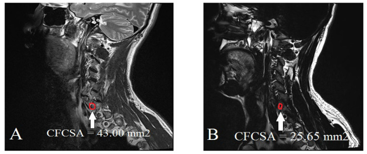
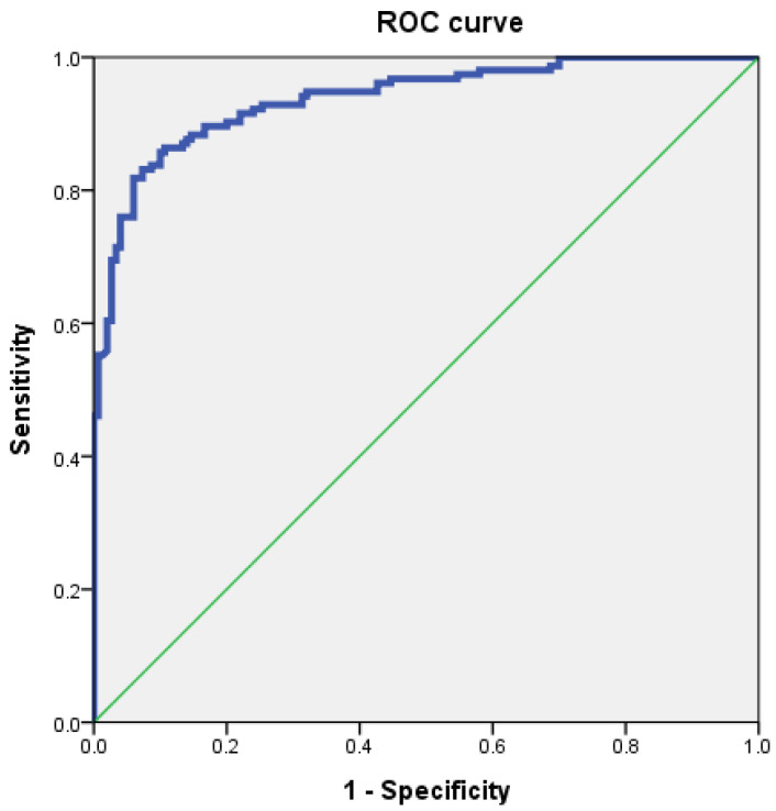
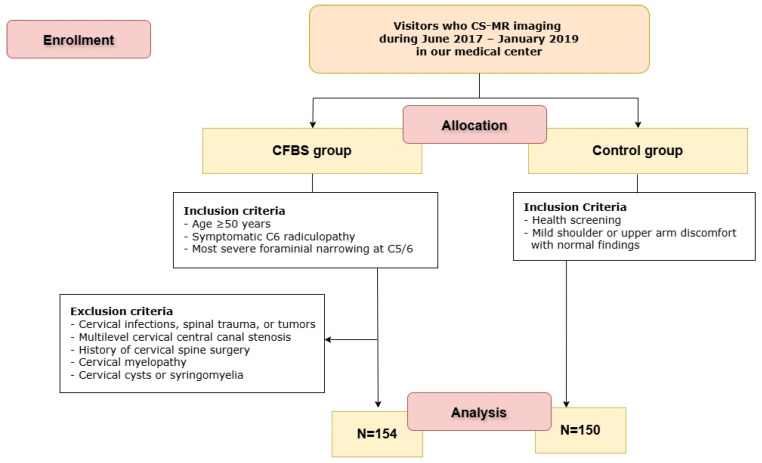
Background: Cervical foraminal bony stenosis (CFBS) is a common degenerative spinal condition that causes radicular pain and functional impairment in the upper extremities. Accurate and objective diagnosis of CFBS remains challenging due to the absence of standardized morphometric criteria. This study aimed to determine an optimal cut-off value for the cervical foraminal cross-sectional area (CFCSA) at the C5/6 level as a diagnostic indicator of CFBS. Methods: We conducted a retrospective case-control study including 154 patients aged 50 years or older with clinically and radiologically confirmed CFBS and 150 age-matched asymptomatic controls. Cervical spine magnetic resonance imaging (MRI) was performed in all subjects and CFCSA measurements were obtained from sagittal T2-weighted images using a standardized protocol. Group differences were analyzed using t-tests and diagnostic performance was assessed using receiver operating characteristic (ROC) curve analysis. Results: The mean CFCSA was significantly lower in the CFBS group (25.65 ± 7.19 mm2) compared to the control group (43.00 ± 8.38 mm2; p < 0.001). ROC analysis identified a CFCSA threshold of 33.02 mm2 as the optimal cut-off point for predicting CFBS, yielding a sensitivity of 86.4%, a specificity of 86.7%, and an area under the curve (AUC) of 0.94 (95% CI: 0.91-0.96). Conclusions: These findings suggest that CFCSA is a robust and reproducible morphological parameter for evaluating foraminal stenosis. The proposed cut-off may enhance diagnostic accuracy and aid in clinical decision-making for patients presenting with C6 radiculopathy. However, given this study's retrospective, single-center design, further validation through multicenter, prospective studies across multiple cervical levels is warranted.
TomographyMedicine-Radiology, Nuclear Medicine and Imaging
CiteScore
2.70
自引率
10.50%
发文量
222
期刊介绍:
TomographyTM publishes basic (technical and pre-clinical) and clinical scientific articles which involve the advancement of imaging technologies. Tomography encompasses studies that use single or multiple imaging modalities including for example CT, US, PET, SPECT, MR and hyperpolarization technologies, as well as optical modalities (i.e. bioluminescence, photoacoustic, endomicroscopy, fiber optic imaging and optical computed tomography) in basic sciences, engineering, preclinical and clinical medicine.
Tomography also welcomes studies involving exploration and refinement of contrast mechanisms and image-derived metrics within and across modalities toward the development of novel imaging probes for image-based feedback and intervention. The use of imaging in biology and medicine provides unparalleled opportunities to noninvasively interrogate tissues to obtain real-time dynamic and quantitative information required for diagnosis and response to interventions and to follow evolving pathological conditions. As multi-modal studies and the complexities of imaging technologies themselves are ever increasing to provide advanced information to scientists and clinicians.
Tomography provides a unique publication venue allowing investigators the opportunity to more precisely communicate integrated findings related to the diverse and heterogeneous features associated with underlying anatomical, physiological, functional, metabolic and molecular genetic activities of normal and diseased tissue. Thus Tomography publishes peer-reviewed articles which involve the broad use of imaging of any tissue and disease type including both preclinical and clinical investigations. In addition, hardware/software along with chemical and molecular probe advances are welcome as they are deemed to significantly contribute towards the long-term goal of improving the overall impact of imaging on scientific and clinical discovery.

 求助内容:
求助内容: 应助结果提醒方式:


