Noor UlAin, Rehan Moinuddin Shaikh, Tayyaba Gul Malik
{"title":"利用椎间盘损伤似然尺度和光学相干断层扫描研究早期和晚期原发性开角型青光眼RNFL损伤模式。","authors":"Noor UlAin, Rehan Moinuddin Shaikh, Tayyaba Gul Malik","doi":"10.4274/tjo.galenos.2025.88834","DOIUrl":null,"url":null,"abstract":"<p><strong>Objectives: </strong>To determine patterns of peripapillary retinal nerve fiber layer (RNFL) damage in early- and late-stage glaucoma based on the Disc Damage Likelihood Scale (DDLS).</p><p><strong>Materials and methods: </strong>This cross-sectional, multi-center study involved 267 eyes of 135 patients aged 18 years or older with suspected or diagnosed glaucoma. Exclusion criteria were high refractive errors, media opacities, trauma history, and systemic conditions affecting the optic disc. After a comprehensive ocular examination, the DDLS was used for glaucoma staging. Disease severity was classified into three zones: green, orange, and red. RNFL thickness was measured in four quadrants using optical coherence tomography. Patterns of RNFL damage were analyzed, especially in terms of the ISNT (inferior>superior>nasal>temporal) rule, and compared between the three groups.</p><p><strong>Results: </strong>The male-to-female ratio was 1.59:1 and the mean age was 45.12±15.76 years. There were statistically significant differences among the groups for average, inferior, superior, and temporal RNFL thickness (p<0.00001). However, the difference in nasal RNFL was insignificant. The ISNT rule was the commonest pattern in the study participants (64.4%) and progressive loss of pattern was observed with increased disease severity.</p><p><strong>Conclusion: </strong>This study revealed an association between disease severity and RNFL thinning in the inferior, superior, and temporal quadrants, while nasal RNFL showed no significant association with disease severity. The ISNT rule was more frequently observed in the early stages and diminished with advanced glaucoma. These results highlight RNFL thinning based on the DDLS as an important marker for glaucoma monitoring.</p>","PeriodicalId":23373,"journal":{"name":"Turkish Journal of Ophthalmology","volume":"55 3","pages":"127-133"},"PeriodicalIF":0.0000,"publicationDate":"2025-06-25","publicationTypes":"Journal Article","fieldsOfStudy":null,"isOpenAccess":false,"openAccessPdf":"https://www.ncbi.nlm.nih.gov/pmc/articles/PMC12192247/pdf/","citationCount":"0","resultStr":"{\"title\":\"Pattern of RNFL Damage in Early- and Late-Stage Primary Open-Angle Glaucoma Using the Disc Damage Likelihood Scale and Optical Coherence Tomography.\",\"authors\":\"Noor UlAin, Rehan Moinuddin Shaikh, Tayyaba Gul Malik\",\"doi\":\"10.4274/tjo.galenos.2025.88834\",\"DOIUrl\":null,\"url\":null,\"abstract\":\"<p><strong>Objectives: </strong>To determine patterns of peripapillary retinal nerve fiber layer (RNFL) damage in early- and late-stage glaucoma based on the Disc Damage Likelihood Scale (DDLS).</p><p><strong>Materials and methods: </strong>This cross-sectional, multi-center study involved 267 eyes of 135 patients aged 18 years or older with suspected or diagnosed glaucoma. Exclusion criteria were high refractive errors, media opacities, trauma history, and systemic conditions affecting the optic disc. After a comprehensive ocular examination, the DDLS was used for glaucoma staging. Disease severity was classified into three zones: green, orange, and red. RNFL thickness was measured in four quadrants using optical coherence tomography. Patterns of RNFL damage were analyzed, especially in terms of the ISNT (inferior>superior>nasal>temporal) rule, and compared between the three groups.</p><p><strong>Results: </strong>The male-to-female ratio was 1.59:1 and the mean age was 45.12±15.76 years. There were statistically significant differences among the groups for average, inferior, superior, and temporal RNFL thickness (p<0.00001). However, the difference in nasal RNFL was insignificant. The ISNT rule was the commonest pattern in the study participants (64.4%) and progressive loss of pattern was observed with increased disease severity.</p><p><strong>Conclusion: </strong>This study revealed an association between disease severity and RNFL thinning in the inferior, superior, and temporal quadrants, while nasal RNFL showed no significant association with disease severity. The ISNT rule was more frequently observed in the early stages and diminished with advanced glaucoma. These results highlight RNFL thinning based on the DDLS as an important marker for glaucoma monitoring.</p>\",\"PeriodicalId\":23373,\"journal\":{\"name\":\"Turkish Journal of Ophthalmology\",\"volume\":\"55 3\",\"pages\":\"127-133\"},\"PeriodicalIF\":0.0000,\"publicationDate\":\"2025-06-25\",\"publicationTypes\":\"Journal Article\",\"fieldsOfStudy\":null,\"isOpenAccess\":false,\"openAccessPdf\":\"https://www.ncbi.nlm.nih.gov/pmc/articles/PMC12192247/pdf/\",\"citationCount\":\"0\",\"resultStr\":null,\"platform\":\"Semanticscholar\",\"paperid\":null,\"PeriodicalName\":\"Turkish Journal of Ophthalmology\",\"FirstCategoryId\":\"1085\",\"ListUrlMain\":\"https://doi.org/10.4274/tjo.galenos.2025.88834\",\"RegionNum\":0,\"RegionCategory\":null,\"ArticlePicture\":[],\"TitleCN\":null,\"AbstractTextCN\":null,\"PMCID\":null,\"EPubDate\":\"\",\"PubModel\":\"\",\"JCR\":\"Q3\",\"JCRName\":\"Medicine\",\"Score\":null,\"Total\":0}","platform":"Semanticscholar","paperid":null,"PeriodicalName":"Turkish Journal of Ophthalmology","FirstCategoryId":"1085","ListUrlMain":"https://doi.org/10.4274/tjo.galenos.2025.88834","RegionNum":0,"RegionCategory":null,"ArticlePicture":[],"TitleCN":null,"AbstractTextCN":null,"PMCID":null,"EPubDate":"","PubModel":"","JCR":"Q3","JCRName":"Medicine","Score":null,"Total":0}
Pattern of RNFL Damage in Early- and Late-Stage Primary Open-Angle Glaucoma Using the Disc Damage Likelihood Scale and Optical Coherence Tomography.
Objectives: To determine patterns of peripapillary retinal nerve fiber layer (RNFL) damage in early- and late-stage glaucoma based on the Disc Damage Likelihood Scale (DDLS).
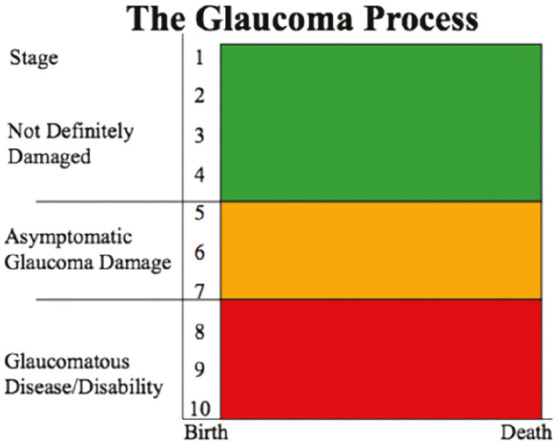
Materials and methods: This cross-sectional, multi-center study involved 267 eyes of 135 patients aged 18 years or older with suspected or diagnosed glaucoma. Exclusion criteria were high refractive errors, media opacities, trauma history, and systemic conditions affecting the optic disc. After a comprehensive ocular examination, the DDLS was used for glaucoma staging. Disease severity was classified into three zones: green, orange, and red. RNFL thickness was measured in four quadrants using optical coherence tomography. Patterns of RNFL damage were analyzed, especially in terms of the ISNT (inferior>superior>nasal>temporal) rule, and compared between the three groups.
Results: The male-to-female ratio was 1.59:1 and the mean age was 45.12±15.76 years. There were statistically significant differences among the groups for average, inferior, superior, and temporal RNFL thickness (p<0.00001). However, the difference in nasal RNFL was insignificant. The ISNT rule was the commonest pattern in the study participants (64.4%) and progressive loss of pattern was observed with increased disease severity.
Conclusion: This study revealed an association between disease severity and RNFL thinning in the inferior, superior, and temporal quadrants, while nasal RNFL showed no significant association with disease severity. The ISNT rule was more frequently observed in the early stages and diminished with advanced glaucoma. These results highlight RNFL thinning based on the DDLS as an important marker for glaucoma monitoring.
期刊介绍:
The Turkish Journal of Ophthalmology (TJO) is the only scientific periodical publication of the Turkish Ophthalmological Association and has been published since January 1929. In its early years, the journal was published in Turkish and French. Although there were temporary interruptions in the publication of the journal due to various challenges, the Turkish Journal of Ophthalmology has been published continually from 1971 to the present. The target audience includes specialists and physicians in training in ophthalmology in all relevant disciplines.

 求助内容:
求助内容: 应助结果提醒方式:


