Andrew P Robinson, Kelley M Ferreira, Warda Heetun, Manuel Bardiès, Ana M Denis-Bacelar, Andrew J Fenwick, Michael Lassmann, Jill Tipping, Johannes Tran-Gia
{"title":"建立定量SPECT成像的测量溯源性。","authors":"Andrew P Robinson, Kelley M Ferreira, Warda Heetun, Manuel Bardiès, Ana M Denis-Bacelar, Andrew J Fenwick, Michael Lassmann, Jill Tipping, Johannes Tran-Gia","doi":"10.1186/s40658-025-00778-9","DOIUrl":null,"url":null,"abstract":"<p><strong>Background: </strong>Single Photon Emission Computed Tomography (SPECT) is increasingly used as a quantitative modality, especially in the context of Molecular Radiotherapy, where the measurements are used as input to absorbed dose calculations for patient-specific dosimetry. Establishing measurement traceability is an essential step in providing confidence in quantitative measurements. This requires an unbroken chain of calibrations where uncertainties must be reported in all stages of calibration and for the final measurement result. Traceability ensures that a measurement result can be related to an underlying standard, allowing harmonisation of data, and facilitating comparison of results between sites.</p><p><strong>Methods: </strong>The process of establishing measurement traceability for quantitative SPECT is demonstrated for the therapeutic radionuclide <sup>177</sup>Lu using a common, phantom based, calibration method. Phantoms with activities of <sup>177</sup>Lu, measured using a traceably calibrated radionuclide calibrator, were used to perform the calibration. The calibration was validated using 3D-printed anthropomorphic organ phantom inserts mimicking clinically relevant geometries. For all measurements, traceability to primary standards for radioactivity is demonstrated along with an accompanying calibration chain and statement of uncertainty.</p><p><strong>Results: </strong>For all activity measurements the dominant component in the activity uncertainty budget was the uncertainty on the radionuclide calibrator calibration factor, resulting in an average combined standard uncertainty of 1.57%. The resulting uncertainty on the SPECT Image Calibration Factor was 1.6%. An optional additional correction was included in the calibration to provide volume-based partial volume correction (PVC). Measurement traceability was extended for measurands using this additional correction. The activity recovery in the organ phantoms with PVC applied was 96(7)% for both the kidney and spleen.</p><p><strong>Conclusions: </strong>A manufacturer independent methodology for establishing measurement traceability for quantitative SPECT is demonstrated for <sup>177</sup>Lu, using a radionuclide calibrator previously calibrated against national standards. The ability to establish measurement traceability for quantitative SPECT using standard clinical equipment, and the limitations of traceability are presented.</p>","PeriodicalId":11559,"journal":{"name":"EJNMMI Physics","volume":"12 1","pages":"58"},"PeriodicalIF":3.2000,"publicationDate":"2025-06-23","publicationTypes":"Journal Article","fieldsOfStudy":null,"isOpenAccess":false,"openAccessPdf":"https://www.ncbi.nlm.nih.gov/pmc/articles/PMC12185843/pdf/","citationCount":"0","resultStr":"{\"title\":\"Establishing measurement traceability for quantitative SPECT imaging.\",\"authors\":\"Andrew P Robinson, Kelley M Ferreira, Warda Heetun, Manuel Bardiès, Ana M Denis-Bacelar, Andrew J Fenwick, Michael Lassmann, Jill Tipping, Johannes Tran-Gia\",\"doi\":\"10.1186/s40658-025-00778-9\",\"DOIUrl\":null,\"url\":null,\"abstract\":\"<p><strong>Background: </strong>Single Photon Emission Computed Tomography (SPECT) is increasingly used as a quantitative modality, especially in the context of Molecular Radiotherapy, where the measurements are used as input to absorbed dose calculations for patient-specific dosimetry. Establishing measurement traceability is an essential step in providing confidence in quantitative measurements. This requires an unbroken chain of calibrations where uncertainties must be reported in all stages of calibration and for the final measurement result. Traceability ensures that a measurement result can be related to an underlying standard, allowing harmonisation of data, and facilitating comparison of results between sites.</p><p><strong>Methods: </strong>The process of establishing measurement traceability for quantitative SPECT is demonstrated for the therapeutic radionuclide <sup>177</sup>Lu using a common, phantom based, calibration method. Phantoms with activities of <sup>177</sup>Lu, measured using a traceably calibrated radionuclide calibrator, were used to perform the calibration. The calibration was validated using 3D-printed anthropomorphic organ phantom inserts mimicking clinically relevant geometries. For all measurements, traceability to primary standards for radioactivity is demonstrated along with an accompanying calibration chain and statement of uncertainty.</p><p><strong>Results: </strong>For all activity measurements the dominant component in the activity uncertainty budget was the uncertainty on the radionuclide calibrator calibration factor, resulting in an average combined standard uncertainty of 1.57%. The resulting uncertainty on the SPECT Image Calibration Factor was 1.6%. An optional additional correction was included in the calibration to provide volume-based partial volume correction (PVC). Measurement traceability was extended for measurands using this additional correction. The activity recovery in the organ phantoms with PVC applied was 96(7)% for both the kidney and spleen.</p><p><strong>Conclusions: </strong>A manufacturer independent methodology for establishing measurement traceability for quantitative SPECT is demonstrated for <sup>177</sup>Lu, using a radionuclide calibrator previously calibrated against national standards. The ability to establish measurement traceability for quantitative SPECT using standard clinical equipment, and the limitations of traceability are presented.</p>\",\"PeriodicalId\":11559,\"journal\":{\"name\":\"EJNMMI Physics\",\"volume\":\"12 1\",\"pages\":\"58\"},\"PeriodicalIF\":3.2000,\"publicationDate\":\"2025-06-23\",\"publicationTypes\":\"Journal Article\",\"fieldsOfStudy\":null,\"isOpenAccess\":false,\"openAccessPdf\":\"https://www.ncbi.nlm.nih.gov/pmc/articles/PMC12185843/pdf/\",\"citationCount\":\"0\",\"resultStr\":null,\"platform\":\"Semanticscholar\",\"paperid\":null,\"PeriodicalName\":\"EJNMMI Physics\",\"FirstCategoryId\":\"3\",\"ListUrlMain\":\"https://doi.org/10.1186/s40658-025-00778-9\",\"RegionNum\":2,\"RegionCategory\":\"医学\",\"ArticlePicture\":[],\"TitleCN\":null,\"AbstractTextCN\":null,\"PMCID\":null,\"EPubDate\":\"\",\"PubModel\":\"\",\"JCR\":\"Q2\",\"JCRName\":\"RADIOLOGY, NUCLEAR MEDICINE & MEDICAL IMAGING\",\"Score\":null,\"Total\":0}","platform":"Semanticscholar","paperid":null,"PeriodicalName":"EJNMMI Physics","FirstCategoryId":"3","ListUrlMain":"https://doi.org/10.1186/s40658-025-00778-9","RegionNum":2,"RegionCategory":"医学","ArticlePicture":[],"TitleCN":null,"AbstractTextCN":null,"PMCID":null,"EPubDate":"","PubModel":"","JCR":"Q2","JCRName":"RADIOLOGY, NUCLEAR MEDICINE & MEDICAL IMAGING","Score":null,"Total":0}
Establishing measurement traceability for quantitative SPECT imaging.
Background: Single Photon Emission Computed Tomography (SPECT) is increasingly used as a quantitative modality, especially in the context of Molecular Radiotherapy, where the measurements are used as input to absorbed dose calculations for patient-specific dosimetry. Establishing measurement traceability is an essential step in providing confidence in quantitative measurements. This requires an unbroken chain of calibrations where uncertainties must be reported in all stages of calibration and for the final measurement result. Traceability ensures that a measurement result can be related to an underlying standard, allowing harmonisation of data, and facilitating comparison of results between sites.
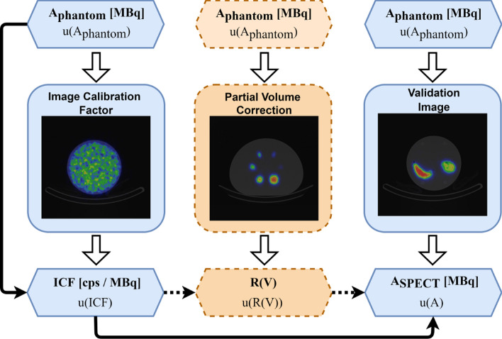
Methods: The process of establishing measurement traceability for quantitative SPECT is demonstrated for the therapeutic radionuclide 177Lu using a common, phantom based, calibration method. Phantoms with activities of 177Lu, measured using a traceably calibrated radionuclide calibrator, were used to perform the calibration. The calibration was validated using 3D-printed anthropomorphic organ phantom inserts mimicking clinically relevant geometries. For all measurements, traceability to primary standards for radioactivity is demonstrated along with an accompanying calibration chain and statement of uncertainty.
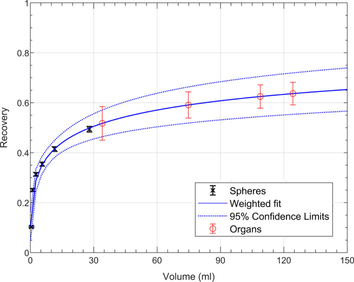
Results: For all activity measurements the dominant component in the activity uncertainty budget was the uncertainty on the radionuclide calibrator calibration factor, resulting in an average combined standard uncertainty of 1.57%. The resulting uncertainty on the SPECT Image Calibration Factor was 1.6%. An optional additional correction was included in the calibration to provide volume-based partial volume correction (PVC). Measurement traceability was extended for measurands using this additional correction. The activity recovery in the organ phantoms with PVC applied was 96(7)% for both the kidney and spleen.
Conclusions: A manufacturer independent methodology for establishing measurement traceability for quantitative SPECT is demonstrated for 177Lu, using a radionuclide calibrator previously calibrated against national standards. The ability to establish measurement traceability for quantitative SPECT using standard clinical equipment, and the limitations of traceability are presented.
期刊介绍:
EJNMMI Physics is an international platform for scientists, users and adopters of nuclear medicine with a particular interest in physics matters. As a companion journal to the European Journal of Nuclear Medicine and Molecular Imaging, this journal has a multi-disciplinary approach and welcomes original materials and studies with a focus on applied physics and mathematics as well as imaging systems engineering and prototyping in nuclear medicine. This includes physics-driven approaches or algorithms supported by physics that foster early clinical adoption of nuclear medicine imaging and therapy.

 求助内容:
求助内容: 应助结果提醒方式:


