Smruti Mahapatra, Vishal N Bhimarasetty, Abdul Rahim, Colin N Curtis, Paul Gulotta, Korak Sarkar
{"title":"3d打印主动脉弓患者特异性模型用于复杂神经介入病例的高级可视化","authors":"Smruti Mahapatra, Vishal N Bhimarasetty, Abdul Rahim, Colin N Curtis, Paul Gulotta, Korak Sarkar","doi":"10.31486/toj.24.0124","DOIUrl":null,"url":null,"abstract":"<p><strong>Background: </strong>Cerebrovascular disease is a leading cause of death and disability worldwide, and endovascular therapies have become a mainstay of treatment for ischemic stroke. However, tortuous anatomy, particularly of the aortic arch, presents formidable treatment challenges by impeding access to intracranial circulation and thus affecting clinical outcomes.</p><p><strong>Methods: </strong>To better understand the challenges of tortuous anatomy, we fabricated 3D-printed models of the aortic arch and major branch vessels based on the imaging of 4 patients.</p><p><strong>Results: </strong>These patient-specific models were realistic representations of the intricate vascular pathways and provided enhanced visualization of the complex vascular structures. The measured diameters of the 3D-fabricated models closely matched the values reported in the literature, confirming the physical accuracy of the models. Creating an individual anatomic model required an average of 4 hours of digital processing and 13.71 hours of 3D printing, with a materials cost of approximately $17.31.</p><p><strong>Conclusion: </strong>3D-printed patient-specific models used for neurointerventional training and preprocedural planning are a valuable tool for managing complex cerebrovascular anatomy. The advanced visualization provided by these models may enhance preparedness and potentially improve ischemic stroke treatment outcomes.</p>","PeriodicalId":47600,"journal":{"name":"Ochsner Journal","volume":"25 2","pages":"92-98"},"PeriodicalIF":1.2000,"publicationDate":"2025-01-01","publicationTypes":"Journal Article","fieldsOfStudy":null,"isOpenAccess":false,"openAccessPdf":"https://www.ncbi.nlm.nih.gov/pmc/articles/PMC12175768/pdf/","citationCount":"0","resultStr":"{\"title\":\"3D-Printed Patient-Specific Models of the Aortic Arch for Advanced Visualization of Complex Neurointerventional Cases.\",\"authors\":\"Smruti Mahapatra, Vishal N Bhimarasetty, Abdul Rahim, Colin N Curtis, Paul Gulotta, Korak Sarkar\",\"doi\":\"10.31486/toj.24.0124\",\"DOIUrl\":null,\"url\":null,\"abstract\":\"<p><strong>Background: </strong>Cerebrovascular disease is a leading cause of death and disability worldwide, and endovascular therapies have become a mainstay of treatment for ischemic stroke. However, tortuous anatomy, particularly of the aortic arch, presents formidable treatment challenges by impeding access to intracranial circulation and thus affecting clinical outcomes.</p><p><strong>Methods: </strong>To better understand the challenges of tortuous anatomy, we fabricated 3D-printed models of the aortic arch and major branch vessels based on the imaging of 4 patients.</p><p><strong>Results: </strong>These patient-specific models were realistic representations of the intricate vascular pathways and provided enhanced visualization of the complex vascular structures. The measured diameters of the 3D-fabricated models closely matched the values reported in the literature, confirming the physical accuracy of the models. Creating an individual anatomic model required an average of 4 hours of digital processing and 13.71 hours of 3D printing, with a materials cost of approximately $17.31.</p><p><strong>Conclusion: </strong>3D-printed patient-specific models used for neurointerventional training and preprocedural planning are a valuable tool for managing complex cerebrovascular anatomy. The advanced visualization provided by these models may enhance preparedness and potentially improve ischemic stroke treatment outcomes.</p>\",\"PeriodicalId\":47600,\"journal\":{\"name\":\"Ochsner Journal\",\"volume\":\"25 2\",\"pages\":\"92-98\"},\"PeriodicalIF\":1.2000,\"publicationDate\":\"2025-01-01\",\"publicationTypes\":\"Journal Article\",\"fieldsOfStudy\":null,\"isOpenAccess\":false,\"openAccessPdf\":\"https://www.ncbi.nlm.nih.gov/pmc/articles/PMC12175768/pdf/\",\"citationCount\":\"0\",\"resultStr\":null,\"platform\":\"Semanticscholar\",\"paperid\":null,\"PeriodicalName\":\"Ochsner Journal\",\"FirstCategoryId\":\"1085\",\"ListUrlMain\":\"https://doi.org/10.31486/toj.24.0124\",\"RegionNum\":0,\"RegionCategory\":null,\"ArticlePicture\":[],\"TitleCN\":null,\"AbstractTextCN\":null,\"PMCID\":null,\"EPubDate\":\"\",\"PubModel\":\"\",\"JCR\":\"Q2\",\"JCRName\":\"MEDICINE, GENERAL & INTERNAL\",\"Score\":null,\"Total\":0}","platform":"Semanticscholar","paperid":null,"PeriodicalName":"Ochsner Journal","FirstCategoryId":"1085","ListUrlMain":"https://doi.org/10.31486/toj.24.0124","RegionNum":0,"RegionCategory":null,"ArticlePicture":[],"TitleCN":null,"AbstractTextCN":null,"PMCID":null,"EPubDate":"","PubModel":"","JCR":"Q2","JCRName":"MEDICINE, GENERAL & INTERNAL","Score":null,"Total":0}
3D-Printed Patient-Specific Models of the Aortic Arch for Advanced Visualization of Complex Neurointerventional Cases.
Background: Cerebrovascular disease is a leading cause of death and disability worldwide, and endovascular therapies have become a mainstay of treatment for ischemic stroke. However, tortuous anatomy, particularly of the aortic arch, presents formidable treatment challenges by impeding access to intracranial circulation and thus affecting clinical outcomes.
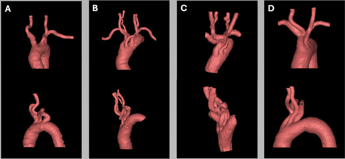
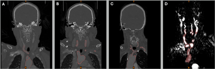
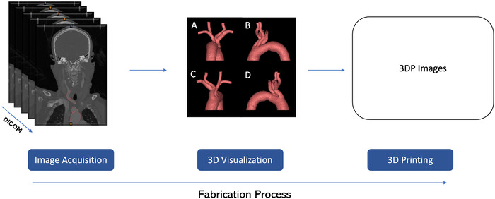
Methods: To better understand the challenges of tortuous anatomy, we fabricated 3D-printed models of the aortic arch and major branch vessels based on the imaging of 4 patients.
Results: These patient-specific models were realistic representations of the intricate vascular pathways and provided enhanced visualization of the complex vascular structures. The measured diameters of the 3D-fabricated models closely matched the values reported in the literature, confirming the physical accuracy of the models. Creating an individual anatomic model required an average of 4 hours of digital processing and 13.71 hours of 3D printing, with a materials cost of approximately $17.31.
Conclusion: 3D-printed patient-specific models used for neurointerventional training and preprocedural planning are a valuable tool for managing complex cerebrovascular anatomy. The advanced visualization provided by these models may enhance preparedness and potentially improve ischemic stroke treatment outcomes.
期刊介绍:
The Ochsner Journal is a quarterly publication designed to support Ochsner"s mission to improve the health of our community through a commitment to innovation in healthcare, medical research, and education. The Ochsner Journal provides an active dialogue on practice standards in today"s changing healthcare environment. Emphasis will be given to topics of great societal and medical significance.

 求助内容:
求助内容: 应助结果提醒方式:


