Noah Mann, Keerthana Surabhi, Josephine Sharp, Mary Phipps, Maelee Becton, Jahiem Hill, Davis Roberts, Erzsebet M Szatmari, Robert M Hughes
{"title":"通过肌动蛋白- atp界面突变鉴定具有神经退行性疾病样表型的肌动蛋白突变体。","authors":"Noah Mann, Keerthana Surabhi, Josephine Sharp, Mary Phipps, Maelee Becton, Jahiem Hill, Davis Roberts, Erzsebet M Szatmari, Robert M Hughes","doi":"10.3389/fncel.2025.1543199","DOIUrl":null,"url":null,"abstract":"<p><p>Cofilin-actin rods are a well-documented stress response in neuronal cells and their persistence is frequently associated with neurodegenerative disease. However, the role of specific actin residues in promoting the formation of cofilin-actin rods and other anomalous cytoskeletal structures is largely unknown. As it is increasingly suspected that specific mutations and post-translation modifications of actin may promote neurodegenerative disease, characterizing the role of these residues in cytoskeletal dysregulation is highly relevant. In this study, we focus on the actin-ATP interface, which has been proposed as a key mediator of cofilin-actin rod formation and the propensity of actin to respond to cellular stress. Using a light and stress-gated reporter of cofilin-actin cluster formation, we determine the impact of mutants associated with Actin-ATP binding on the propensity of actin to form anomalous structures in the presence and absence of applied cellular stress. This study identifies actin mutants that promote anomalous actin inclusions in HeLa cells and characterizes the manifestation of these phenotypes in cortical neurons. Mutations to the ATP phosphate tail-binding region of actin (K18A, D154A, G158L, K213A) were found to be particularly disruptive to actin phenotypes, and in several instances promote disease-associated actin-rich structures such as cofilin-actin rods and Hirano bodies. We find that these mutant phenotypes are largely consistent between cell types and display highly unusual inclusions in cultured cortical neurons, without leading to nuclear fragmentation and apoptotic death of the transfected cells. These mutants strengthen the association of residue-specific changes in actin with large-scale phenotypic and functional changes in the cytoskeleton, further implicating them in neurodegenerative disease progression.</p>","PeriodicalId":12432,"journal":{"name":"Frontiers in Cellular Neuroscience","volume":"19 ","pages":"1543199"},"PeriodicalIF":4.0000,"publicationDate":"2025-06-04","publicationTypes":"Journal Article","fieldsOfStudy":null,"isOpenAccess":false,"openAccessPdf":"https://www.ncbi.nlm.nih.gov/pmc/articles/PMC12174402/pdf/","citationCount":"0","resultStr":"{\"title\":\"Identification of actin mutants with neurodegenerative disease-like phenotypes via mutagenesis of the actin-ATP interface.\",\"authors\":\"Noah Mann, Keerthana Surabhi, Josephine Sharp, Mary Phipps, Maelee Becton, Jahiem Hill, Davis Roberts, Erzsebet M Szatmari, Robert M Hughes\",\"doi\":\"10.3389/fncel.2025.1543199\",\"DOIUrl\":null,\"url\":null,\"abstract\":\"<p><p>Cofilin-actin rods are a well-documented stress response in neuronal cells and their persistence is frequently associated with neurodegenerative disease. However, the role of specific actin residues in promoting the formation of cofilin-actin rods and other anomalous cytoskeletal structures is largely unknown. As it is increasingly suspected that specific mutations and post-translation modifications of actin may promote neurodegenerative disease, characterizing the role of these residues in cytoskeletal dysregulation is highly relevant. In this study, we focus on the actin-ATP interface, which has been proposed as a key mediator of cofilin-actin rod formation and the propensity of actin to respond to cellular stress. Using a light and stress-gated reporter of cofilin-actin cluster formation, we determine the impact of mutants associated with Actin-ATP binding on the propensity of actin to form anomalous structures in the presence and absence of applied cellular stress. This study identifies actin mutants that promote anomalous actin inclusions in HeLa cells and characterizes the manifestation of these phenotypes in cortical neurons. Mutations to the ATP phosphate tail-binding region of actin (K18A, D154A, G158L, K213A) were found to be particularly disruptive to actin phenotypes, and in several instances promote disease-associated actin-rich structures such as cofilin-actin rods and Hirano bodies. We find that these mutant phenotypes are largely consistent between cell types and display highly unusual inclusions in cultured cortical neurons, without leading to nuclear fragmentation and apoptotic death of the transfected cells. These mutants strengthen the association of residue-specific changes in actin with large-scale phenotypic and functional changes in the cytoskeleton, further implicating them in neurodegenerative disease progression.</p>\",\"PeriodicalId\":12432,\"journal\":{\"name\":\"Frontiers in Cellular Neuroscience\",\"volume\":\"19 \",\"pages\":\"1543199\"},\"PeriodicalIF\":4.0000,\"publicationDate\":\"2025-06-04\",\"publicationTypes\":\"Journal Article\",\"fieldsOfStudy\":null,\"isOpenAccess\":false,\"openAccessPdf\":\"https://www.ncbi.nlm.nih.gov/pmc/articles/PMC12174402/pdf/\",\"citationCount\":\"0\",\"resultStr\":null,\"platform\":\"Semanticscholar\",\"paperid\":null,\"PeriodicalName\":\"Frontiers in Cellular Neuroscience\",\"FirstCategoryId\":\"3\",\"ListUrlMain\":\"https://doi.org/10.3389/fncel.2025.1543199\",\"RegionNum\":3,\"RegionCategory\":\"医学\",\"ArticlePicture\":[],\"TitleCN\":null,\"AbstractTextCN\":null,\"PMCID\":null,\"EPubDate\":\"2025/1/1 0:00:00\",\"PubModel\":\"eCollection\",\"JCR\":\"Q2\",\"JCRName\":\"NEUROSCIENCES\",\"Score\":null,\"Total\":0}","platform":"Semanticscholar","paperid":null,"PeriodicalName":"Frontiers in Cellular Neuroscience","FirstCategoryId":"3","ListUrlMain":"https://doi.org/10.3389/fncel.2025.1543199","RegionNum":3,"RegionCategory":"医学","ArticlePicture":[],"TitleCN":null,"AbstractTextCN":null,"PMCID":null,"EPubDate":"2025/1/1 0:00:00","PubModel":"eCollection","JCR":"Q2","JCRName":"NEUROSCIENCES","Score":null,"Total":0}
Identification of actin mutants with neurodegenerative disease-like phenotypes via mutagenesis of the actin-ATP interface.
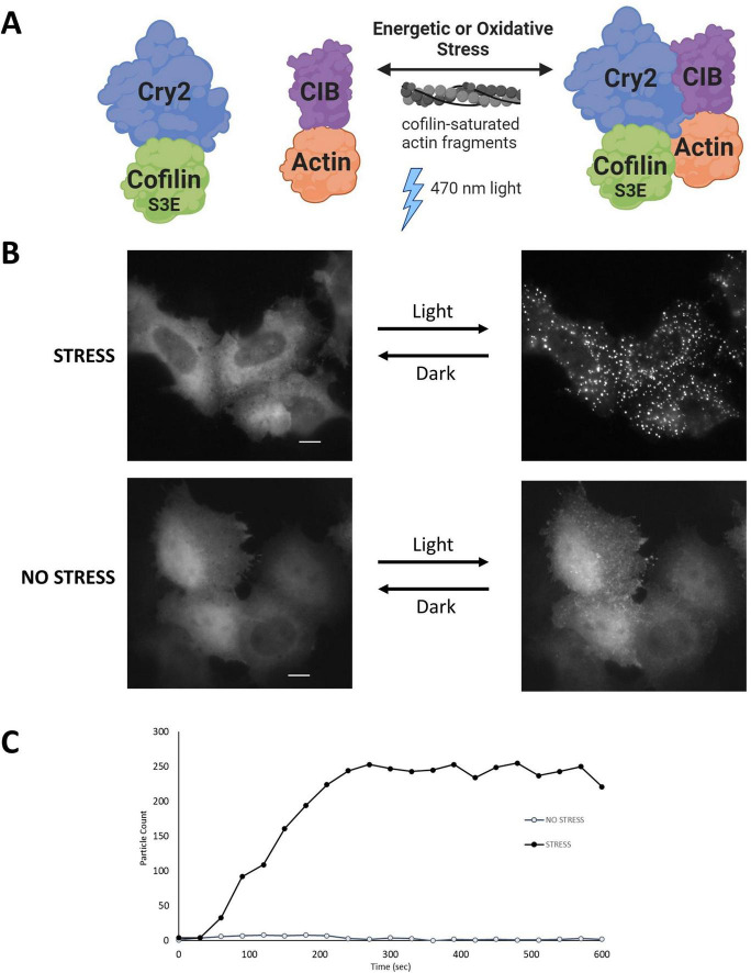
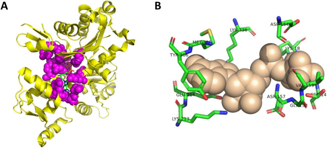
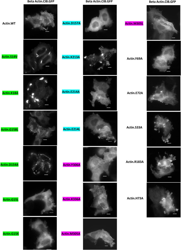
Cofilin-actin rods are a well-documented stress response in neuronal cells and their persistence is frequently associated with neurodegenerative disease. However, the role of specific actin residues in promoting the formation of cofilin-actin rods and other anomalous cytoskeletal structures is largely unknown. As it is increasingly suspected that specific mutations and post-translation modifications of actin may promote neurodegenerative disease, characterizing the role of these residues in cytoskeletal dysregulation is highly relevant. In this study, we focus on the actin-ATP interface, which has been proposed as a key mediator of cofilin-actin rod formation and the propensity of actin to respond to cellular stress. Using a light and stress-gated reporter of cofilin-actin cluster formation, we determine the impact of mutants associated with Actin-ATP binding on the propensity of actin to form anomalous structures in the presence and absence of applied cellular stress. This study identifies actin mutants that promote anomalous actin inclusions in HeLa cells and characterizes the manifestation of these phenotypes in cortical neurons. Mutations to the ATP phosphate tail-binding region of actin (K18A, D154A, G158L, K213A) were found to be particularly disruptive to actin phenotypes, and in several instances promote disease-associated actin-rich structures such as cofilin-actin rods and Hirano bodies. We find that these mutant phenotypes are largely consistent between cell types and display highly unusual inclusions in cultured cortical neurons, without leading to nuclear fragmentation and apoptotic death of the transfected cells. These mutants strengthen the association of residue-specific changes in actin with large-scale phenotypic and functional changes in the cytoskeleton, further implicating them in neurodegenerative disease progression.
期刊介绍:
Frontiers in Cellular Neuroscience is a leading journal in its field, publishing rigorously peer-reviewed research that advances our understanding of the cellular mechanisms underlying cell function in the nervous system across all species. Specialty Chief Editors Egidio D‘Angelo at the University of Pavia and Christian Hansel at the University of Chicago are supported by an outstanding Editorial Board of international researchers. This multidisciplinary open-access journal is at the forefront of disseminating and communicating scientific knowledge and impactful discoveries to researchers, academics, clinicians and the public worldwide.
 求助内容:
求助内容: 应助结果提醒方式:


