Camiel J Smees, Judith Olde Heuvel, Stein van der Heide, Esmee D van Uum, Anne J H Vochteloo, Gabriëlle J M Tuijthof
{"title":"桡骨远端畸形愈合矫正性截骨的形状完成模型。","authors":"Camiel J Smees, Judith Olde Heuvel, Stein van der Heide, Esmee D van Uum, Anne J H Vochteloo, Gabriëlle J M Tuijthof","doi":"10.1007/s11548-025-03454-6","DOIUrl":null,"url":null,"abstract":"<p><strong>Purpose: </strong>When performing 3D planning for osteotomies in patients with distal radius malunion, the contralateral radius is commonly used as a template for reconstruction. However, in approximately 10% of the cases, the contralateral radius is not suitable for use. A shape completion model may provide an alternative by generating a healthy radius model based on the proximal part of the malunited bone. The aim of this study is to develop and clinically evaluate such a shape completion model.</p><p><strong>Method: </strong>A total of 100 segmented CT scans of healthy radii were used, with 80 scans used to train a statistical shape model (SSM). This SSM formed the base for a shape completion model capable of predicting the distal 12% based on the proximal 88%. Hyperparameters were optimized using 10 segmented 3D models, and the remaining 10 models were reserved for testing the performance of the shape completion model.</p><p><strong>Results: </strong>The shape completion model consistently produced clinically viable 3D reconstructions. The mean absolute errors between the predicted and corresponding reference models in the rotational errors were 2.6 ± 1.7° for radial inclination, 3.6 ± 2.2° for volar tilt, and 2.6 ± 2.8° for axial rotation. Translational errors were 0.7 ± 0.6 mm in dorsal shift, 0.8 ± 0.5 mm in radial shift, and 1.7 ± 1.1 mm in lengthening.</p><p><strong>Conclusion: </strong>This study successfully developed a shape completion model capable of reconstructing healthy 3D radius models based on the proximal bone. The observed errors indicate that the model is viable for use in 3D planning for patients lacking a healthy contralateral radius. However, routine use in patients with a healthy contralateral radius is not yet advised, as error margins exceed bilateral differences observed in healthy populations. The most clinically relevant error found in the model, length mismatch, can be easily corrected during 3D planning if the ipsilateral ulna remains intact.</p>","PeriodicalId":51251,"journal":{"name":"International Journal of Computer Assisted Radiology and Surgery","volume":" ","pages":"2075-2085"},"PeriodicalIF":2.3000,"publicationDate":"2025-10-01","publicationTypes":"Journal Article","fieldsOfStudy":null,"isOpenAccess":false,"openAccessPdf":"https://www.ncbi.nlm.nih.gov/pmc/articles/PMC12518486/pdf/","citationCount":"0","resultStr":"{\"title\":\"A shape completion model for corrective osteotomy of distal radius malunion.\",\"authors\":\"Camiel J Smees, Judith Olde Heuvel, Stein van der Heide, Esmee D van Uum, Anne J H Vochteloo, Gabriëlle J M Tuijthof\",\"doi\":\"10.1007/s11548-025-03454-6\",\"DOIUrl\":null,\"url\":null,\"abstract\":\"<p><strong>Purpose: </strong>When performing 3D planning for osteotomies in patients with distal radius malunion, the contralateral radius is commonly used as a template for reconstruction. However, in approximately 10% of the cases, the contralateral radius is not suitable for use. A shape completion model may provide an alternative by generating a healthy radius model based on the proximal part of the malunited bone. The aim of this study is to develop and clinically evaluate such a shape completion model.</p><p><strong>Method: </strong>A total of 100 segmented CT scans of healthy radii were used, with 80 scans used to train a statistical shape model (SSM). This SSM formed the base for a shape completion model capable of predicting the distal 12% based on the proximal 88%. Hyperparameters were optimized using 10 segmented 3D models, and the remaining 10 models were reserved for testing the performance of the shape completion model.</p><p><strong>Results: </strong>The shape completion model consistently produced clinically viable 3D reconstructions. The mean absolute errors between the predicted and corresponding reference models in the rotational errors were 2.6 ± 1.7° for radial inclination, 3.6 ± 2.2° for volar tilt, and 2.6 ± 2.8° for axial rotation. Translational errors were 0.7 ± 0.6 mm in dorsal shift, 0.8 ± 0.5 mm in radial shift, and 1.7 ± 1.1 mm in lengthening.</p><p><strong>Conclusion: </strong>This study successfully developed a shape completion model capable of reconstructing healthy 3D radius models based on the proximal bone. The observed errors indicate that the model is viable for use in 3D planning for patients lacking a healthy contralateral radius. However, routine use in patients with a healthy contralateral radius is not yet advised, as error margins exceed bilateral differences observed in healthy populations. The most clinically relevant error found in the model, length mismatch, can be easily corrected during 3D planning if the ipsilateral ulna remains intact.</p>\",\"PeriodicalId\":51251,\"journal\":{\"name\":\"International Journal of Computer Assisted Radiology and Surgery\",\"volume\":\" \",\"pages\":\"2075-2085\"},\"PeriodicalIF\":2.3000,\"publicationDate\":\"2025-10-01\",\"publicationTypes\":\"Journal Article\",\"fieldsOfStudy\":null,\"isOpenAccess\":false,\"openAccessPdf\":\"https://www.ncbi.nlm.nih.gov/pmc/articles/PMC12518486/pdf/\",\"citationCount\":\"0\",\"resultStr\":null,\"platform\":\"Semanticscholar\",\"paperid\":null,\"PeriodicalName\":\"International Journal of Computer Assisted Radiology and Surgery\",\"FirstCategoryId\":\"5\",\"ListUrlMain\":\"https://doi.org/10.1007/s11548-025-03454-6\",\"RegionNum\":3,\"RegionCategory\":\"医学\",\"ArticlePicture\":[],\"TitleCN\":null,\"AbstractTextCN\":null,\"PMCID\":null,\"EPubDate\":\"2025/6/17 0:00:00\",\"PubModel\":\"Epub\",\"JCR\":\"Q3\",\"JCRName\":\"ENGINEERING, BIOMEDICAL\",\"Score\":null,\"Total\":0}","platform":"Semanticscholar","paperid":null,"PeriodicalName":"International Journal of Computer Assisted Radiology and Surgery","FirstCategoryId":"5","ListUrlMain":"https://doi.org/10.1007/s11548-025-03454-6","RegionNum":3,"RegionCategory":"医学","ArticlePicture":[],"TitleCN":null,"AbstractTextCN":null,"PMCID":null,"EPubDate":"2025/6/17 0:00:00","PubModel":"Epub","JCR":"Q3","JCRName":"ENGINEERING, BIOMEDICAL","Score":null,"Total":0}
A shape completion model for corrective osteotomy of distal radius malunion.
Purpose: When performing 3D planning for osteotomies in patients with distal radius malunion, the contralateral radius is commonly used as a template for reconstruction. However, in approximately 10% of the cases, the contralateral radius is not suitable for use. A shape completion model may provide an alternative by generating a healthy radius model based on the proximal part of the malunited bone. The aim of this study is to develop and clinically evaluate such a shape completion model.
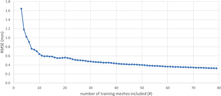
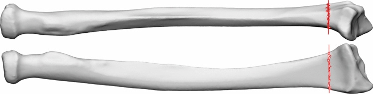
Method: A total of 100 segmented CT scans of healthy radii were used, with 80 scans used to train a statistical shape model (SSM). This SSM formed the base for a shape completion model capable of predicting the distal 12% based on the proximal 88%. Hyperparameters were optimized using 10 segmented 3D models, and the remaining 10 models were reserved for testing the performance of the shape completion model.
Results: The shape completion model consistently produced clinically viable 3D reconstructions. The mean absolute errors between the predicted and corresponding reference models in the rotational errors were 2.6 ± 1.7° for radial inclination, 3.6 ± 2.2° for volar tilt, and 2.6 ± 2.8° for axial rotation. Translational errors were 0.7 ± 0.6 mm in dorsal shift, 0.8 ± 0.5 mm in radial shift, and 1.7 ± 1.1 mm in lengthening.
Conclusion: This study successfully developed a shape completion model capable of reconstructing healthy 3D radius models based on the proximal bone. The observed errors indicate that the model is viable for use in 3D planning for patients lacking a healthy contralateral radius. However, routine use in patients with a healthy contralateral radius is not yet advised, as error margins exceed bilateral differences observed in healthy populations. The most clinically relevant error found in the model, length mismatch, can be easily corrected during 3D planning if the ipsilateral ulna remains intact.
期刊介绍:
The International Journal for Computer Assisted Radiology and Surgery (IJCARS) is a peer-reviewed journal that provides a platform for closing the gap between medical and technical disciplines, and encourages interdisciplinary research and development activities in an international environment.


 求助内容:
求助内容: 应助结果提醒方式:


