Harshmeet Kaur, Rajni Kudawla, Tanmay Pandey and Tripta Bhatia*,
{"title":"实验确定的质膜囊泡,磷脂酰胆碱(PC)和PC-胆固醇囊泡的形状:用共聚焦显微镜进行囊泡收缩分析","authors":"Harshmeet Kaur, Rajni Kudawla, Tanmay Pandey and Tripta Bhatia*, ","doi":"10.1021/acs.jpcb.4c0743110.1021/acs.jpcb.4c07431","DOIUrl":null,"url":null,"abstract":"<p >Giant membrane vesicles (GUVs) and giant plasma membrane vesicles (GPMVs) are valuable models for studying the properties of cellular membranes. We analyzed experimental data on vesicle shapes in three-dimensional space to estimate their reduced volumes, focusing on osmotic deflation and membrane asymmetry. Shape changes in GPMVs illustrate how osmolarity influences the membrane structure in the absence of the cytoskeleton or other cellular organelles. By examining the experimentally observed shapes and their corresponding reduced volumes, we compared GPMV shapes to theoretical predictions for simple phospholipid vesicles, utilizing the area-difference elasticity and spontaneous curvature models. We mapped DOPC GUVs using the area-difference elasticity model and applied the spontaneous curvature model to map DOPC: cholesterol GUVs and GPMVs. The reported experiments showcase advanced methods that provide valuable biophysical insights, demonstrating that the GPMV shape observed in the experiments and their reduced volume can be mapped onto the same shape diagram as red blood cells (RBCs) and vesicles composed of phospholipids. This finding offers new perspectives in the field.</p>","PeriodicalId":60,"journal":{"name":"The Journal of Physical Chemistry B","volume":"129 24","pages":"5913–5922 5913–5922"},"PeriodicalIF":2.9000,"publicationDate":"2025-06-05","publicationTypes":"Journal Article","fieldsOfStudy":null,"isOpenAccess":false,"openAccessPdf":"","citationCount":"0","resultStr":"{\"title\":\"Experimentally Determined Shapes of Plasma Membrane Vesicles, Phosphatidylcholine (PC), and PC-Cholesterol Vesicles: Vesicle Deflation Analysis Using Confocal Microscopy\",\"authors\":\"Harshmeet Kaur, Rajni Kudawla, Tanmay Pandey and Tripta Bhatia*, \",\"doi\":\"10.1021/acs.jpcb.4c0743110.1021/acs.jpcb.4c07431\",\"DOIUrl\":null,\"url\":null,\"abstract\":\"<p >Giant membrane vesicles (GUVs) and giant plasma membrane vesicles (GPMVs) are valuable models for studying the properties of cellular membranes. We analyzed experimental data on vesicle shapes in three-dimensional space to estimate their reduced volumes, focusing on osmotic deflation and membrane asymmetry. Shape changes in GPMVs illustrate how osmolarity influences the membrane structure in the absence of the cytoskeleton or other cellular organelles. By examining the experimentally observed shapes and their corresponding reduced volumes, we compared GPMV shapes to theoretical predictions for simple phospholipid vesicles, utilizing the area-difference elasticity and spontaneous curvature models. We mapped DOPC GUVs using the area-difference elasticity model and applied the spontaneous curvature model to map DOPC: cholesterol GUVs and GPMVs. The reported experiments showcase advanced methods that provide valuable biophysical insights, demonstrating that the GPMV shape observed in the experiments and their reduced volume can be mapped onto the same shape diagram as red blood cells (RBCs) and vesicles composed of phospholipids. This finding offers new perspectives in the field.</p>\",\"PeriodicalId\":60,\"journal\":{\"name\":\"The Journal of Physical Chemistry B\",\"volume\":\"129 24\",\"pages\":\"5913–5922 5913–5922\"},\"PeriodicalIF\":2.9000,\"publicationDate\":\"2025-06-05\",\"publicationTypes\":\"Journal Article\",\"fieldsOfStudy\":null,\"isOpenAccess\":false,\"openAccessPdf\":\"\",\"citationCount\":\"0\",\"resultStr\":null,\"platform\":\"Semanticscholar\",\"paperid\":null,\"PeriodicalName\":\"The Journal of Physical Chemistry B\",\"FirstCategoryId\":\"1\",\"ListUrlMain\":\"https://pubs.acs.org/doi/10.1021/acs.jpcb.4c07431\",\"RegionNum\":2,\"RegionCategory\":\"化学\",\"ArticlePicture\":[],\"TitleCN\":null,\"AbstractTextCN\":null,\"PMCID\":null,\"EPubDate\":\"\",\"PubModel\":\"\",\"JCR\":\"Q3\",\"JCRName\":\"CHEMISTRY, PHYSICAL\",\"Score\":null,\"Total\":0}","platform":"Semanticscholar","paperid":null,"PeriodicalName":"The Journal of Physical Chemistry B","FirstCategoryId":"1","ListUrlMain":"https://pubs.acs.org/doi/10.1021/acs.jpcb.4c07431","RegionNum":2,"RegionCategory":"化学","ArticlePicture":[],"TitleCN":null,"AbstractTextCN":null,"PMCID":null,"EPubDate":"","PubModel":"","JCR":"Q3","JCRName":"CHEMISTRY, PHYSICAL","Score":null,"Total":0}
Experimentally Determined Shapes of Plasma Membrane Vesicles, Phosphatidylcholine (PC), and PC-Cholesterol Vesicles: Vesicle Deflation Analysis Using Confocal Microscopy
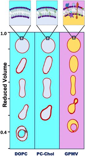
Giant membrane vesicles (GUVs) and giant plasma membrane vesicles (GPMVs) are valuable models for studying the properties of cellular membranes. We analyzed experimental data on vesicle shapes in three-dimensional space to estimate their reduced volumes, focusing on osmotic deflation and membrane asymmetry. Shape changes in GPMVs illustrate how osmolarity influences the membrane structure in the absence of the cytoskeleton or other cellular organelles. By examining the experimentally observed shapes and their corresponding reduced volumes, we compared GPMV shapes to theoretical predictions for simple phospholipid vesicles, utilizing the area-difference elasticity and spontaneous curvature models. We mapped DOPC GUVs using the area-difference elasticity model and applied the spontaneous curvature model to map DOPC: cholesterol GUVs and GPMVs. The reported experiments showcase advanced methods that provide valuable biophysical insights, demonstrating that the GPMV shape observed in the experiments and their reduced volume can be mapped onto the same shape diagram as red blood cells (RBCs) and vesicles composed of phospholipids. This finding offers new perspectives in the field.
期刊介绍:
An essential criterion for acceptance of research articles in the journal is that they provide new physical insight. Please refer to the New Physical Insights virtual issue on what constitutes new physical insight. Manuscripts that are essentially reporting data or applications of data are, in general, not suitable for publication in JPC B.
 求助内容:
求助内容: 应助结果提醒方式:


