Carrie Richardson, Mehrbod S Javadi, Ami A Shah, Caoilfhionn Connolly, Lilja B Solnes, Fredrick M Wigley, Laura K Hummers, Lisa Christopher-Stine
{"title":"18F-NaF PET/CT可识别皮肌炎和系统性硬化症相关钙化症的肌肉和皮下钙化。","authors":"Carrie Richardson, Mehrbod S Javadi, Ami A Shah, Caoilfhionn Connolly, Lilja B Solnes, Fredrick M Wigley, Laura K Hummers, Lisa Christopher-Stine","doi":"10.3389/fnume.2025.1593825","DOIUrl":null,"url":null,"abstract":"<p><strong>Background: </strong>Calcinosis is a morbid complication of dermatomyositis (DM) and systemic sclerosis (SSc) with no effective pharmacologic treatment or validated whole-body assessment modality. <sup>18</sup>F-NaF PET/CT may help to quantify and characterize calcinosis.</p><p><strong>Methods: </strong>In this pilot study, we enrolled three adults with DM and three with SSc, all with new calcinosis deposits. Each underwent <sup>18</sup>F-NaF PET/CT and clinical examination with semi-quantitative scoring of calcinosis. We described the <sup>18</sup>F-NaF PET/CT findings and compared these to CT imaging alone as well as to clinical examination.</p><p><strong>Results: </strong>Calcinosis was noted on <sup>18</sup>F-NaF PET/CT in the subcutaneous tissue in all patients and the muscle in three patients, including two with SSc. The average semi-quantitative score was 23.5 by <sup>18</sup>F-NaF PET/CT and 20 by clinical exam. Wilcoxon signed rank test indicated greater scores by <sup>18</sup>F-NaF PET/CT than by clinical exam (<i>p</i> = 0.0264). <sup>18</sup>F-NaF uptake varied among calcinosis deposits and occurred without corresponding calcifications on CT.</p><p><strong>Conclusions: </strong><sup>18</sup>F-NaF PET/CT appears to be a sensitive method of detecting and characterizing calcinosis that provides both quantitative and qualitative data beyond what can be obtained by physical examination or CT alone. <sup>18</sup>F-NaF uptake occurs in muscle in both SSc and DM, suggesting the possibility that myositis may be driving calcinosis in a subset of patients with SSc.</p>","PeriodicalId":73095,"journal":{"name":"Frontiers in nuclear medicine (Lausanne, Switzerland)","volume":"5 ","pages":"1593825"},"PeriodicalIF":1.4000,"publicationDate":"2025-05-30","publicationTypes":"Journal Article","fieldsOfStudy":null,"isOpenAccess":false,"openAccessPdf":"https://www.ncbi.nlm.nih.gov/pmc/articles/PMC12162990/pdf/","citationCount":"0","resultStr":"{\"title\":\"<sup>18</sup>F-NaF PET/CT identifies muscular and subcutaneous calcifications in both dermatomyositis- and systemic sclerosis-related calcinosis.\",\"authors\":\"Carrie Richardson, Mehrbod S Javadi, Ami A Shah, Caoilfhionn Connolly, Lilja B Solnes, Fredrick M Wigley, Laura K Hummers, Lisa Christopher-Stine\",\"doi\":\"10.3389/fnume.2025.1593825\",\"DOIUrl\":null,\"url\":null,\"abstract\":\"<p><strong>Background: </strong>Calcinosis is a morbid complication of dermatomyositis (DM) and systemic sclerosis (SSc) with no effective pharmacologic treatment or validated whole-body assessment modality. <sup>18</sup>F-NaF PET/CT may help to quantify and characterize calcinosis.</p><p><strong>Methods: </strong>In this pilot study, we enrolled three adults with DM and three with SSc, all with new calcinosis deposits. Each underwent <sup>18</sup>F-NaF PET/CT and clinical examination with semi-quantitative scoring of calcinosis. We described the <sup>18</sup>F-NaF PET/CT findings and compared these to CT imaging alone as well as to clinical examination.</p><p><strong>Results: </strong>Calcinosis was noted on <sup>18</sup>F-NaF PET/CT in the subcutaneous tissue in all patients and the muscle in three patients, including two with SSc. The average semi-quantitative score was 23.5 by <sup>18</sup>F-NaF PET/CT and 20 by clinical exam. Wilcoxon signed rank test indicated greater scores by <sup>18</sup>F-NaF PET/CT than by clinical exam (<i>p</i> = 0.0264). <sup>18</sup>F-NaF uptake varied among calcinosis deposits and occurred without corresponding calcifications on CT.</p><p><strong>Conclusions: </strong><sup>18</sup>F-NaF PET/CT appears to be a sensitive method of detecting and characterizing calcinosis that provides both quantitative and qualitative data beyond what can be obtained by physical examination or CT alone. <sup>18</sup>F-NaF uptake occurs in muscle in both SSc and DM, suggesting the possibility that myositis may be driving calcinosis in a subset of patients with SSc.</p>\",\"PeriodicalId\":73095,\"journal\":{\"name\":\"Frontiers in nuclear medicine (Lausanne, Switzerland)\",\"volume\":\"5 \",\"pages\":\"1593825\"},\"PeriodicalIF\":1.4000,\"publicationDate\":\"2025-05-30\",\"publicationTypes\":\"Journal Article\",\"fieldsOfStudy\":null,\"isOpenAccess\":false,\"openAccessPdf\":\"https://www.ncbi.nlm.nih.gov/pmc/articles/PMC12162990/pdf/\",\"citationCount\":\"0\",\"resultStr\":null,\"platform\":\"Semanticscholar\",\"paperid\":null,\"PeriodicalName\":\"Frontiers in nuclear medicine (Lausanne, Switzerland)\",\"FirstCategoryId\":\"1085\",\"ListUrlMain\":\"https://doi.org/10.3389/fnume.2025.1593825\",\"RegionNum\":0,\"RegionCategory\":null,\"ArticlePicture\":[],\"TitleCN\":null,\"AbstractTextCN\":null,\"PMCID\":null,\"EPubDate\":\"2025/1/1 0:00:00\",\"PubModel\":\"eCollection\",\"JCR\":\"\",\"JCRName\":\"\",\"Score\":null,\"Total\":0}","platform":"Semanticscholar","paperid":null,"PeriodicalName":"Frontiers in nuclear medicine (Lausanne, Switzerland)","FirstCategoryId":"1085","ListUrlMain":"https://doi.org/10.3389/fnume.2025.1593825","RegionNum":0,"RegionCategory":null,"ArticlePicture":[],"TitleCN":null,"AbstractTextCN":null,"PMCID":null,"EPubDate":"2025/1/1 0:00:00","PubModel":"eCollection","JCR":"","JCRName":"","Score":null,"Total":0}
18F-NaF PET/CT identifies muscular and subcutaneous calcifications in both dermatomyositis- and systemic sclerosis-related calcinosis.
Background: Calcinosis is a morbid complication of dermatomyositis (DM) and systemic sclerosis (SSc) with no effective pharmacologic treatment or validated whole-body assessment modality. 18F-NaF PET/CT may help to quantify and characterize calcinosis.
Methods: In this pilot study, we enrolled three adults with DM and three with SSc, all with new calcinosis deposits. Each underwent 18F-NaF PET/CT and clinical examination with semi-quantitative scoring of calcinosis. We described the 18F-NaF PET/CT findings and compared these to CT imaging alone as well as to clinical examination.
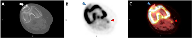
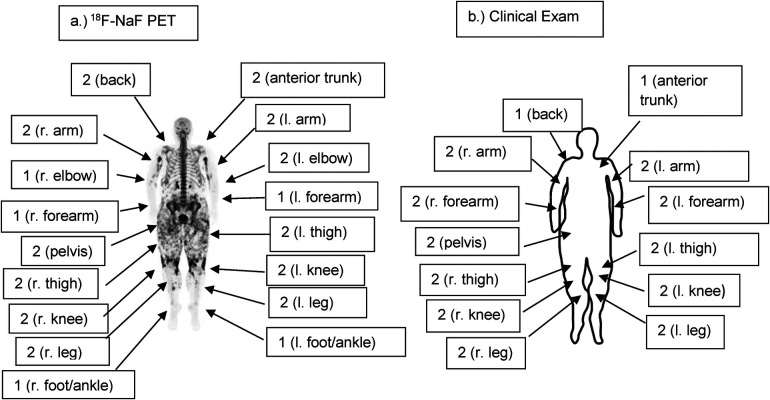
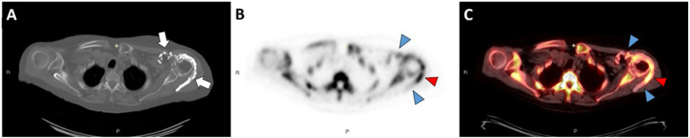
Results: Calcinosis was noted on 18F-NaF PET/CT in the subcutaneous tissue in all patients and the muscle in three patients, including two with SSc. The average semi-quantitative score was 23.5 by 18F-NaF PET/CT and 20 by clinical exam. Wilcoxon signed rank test indicated greater scores by 18F-NaF PET/CT than by clinical exam (p = 0.0264). 18F-NaF uptake varied among calcinosis deposits and occurred without corresponding calcifications on CT.
Conclusions: 18F-NaF PET/CT appears to be a sensitive method of detecting and characterizing calcinosis that provides both quantitative and qualitative data beyond what can be obtained by physical examination or CT alone. 18F-NaF uptake occurs in muscle in both SSc and DM, suggesting the possibility that myositis may be driving calcinosis in a subset of patients with SSc.

 求助内容:
求助内容: 应助结果提醒方式:


