Walter Coudyzer, Lesley Cockmartin, Bram Miseur, Tim Busselot, Didier Bielen, Dirk Vanbeckevoort, Raymond Oyen, Hilde Bosmans
{"title":"个性化腹部CT造影剂体积:理论与实践的桥梁。","authors":"Walter Coudyzer, Lesley Cockmartin, Bram Miseur, Tim Busselot, Didier Bielen, Dirk Vanbeckevoort, Raymond Oyen, Hilde Bosmans","doi":"10.5334/jbsr.3906","DOIUrl":null,"url":null,"abstract":"<p><p><i>Objectives:</i> to determine (1) a target Hounsfield unit (HU) for portal venous phase iodinated contrast media enhanced abdominal CT scans, (2) an equation for a personalized total contrast agent volume, and (3) the best/most appropriate time between injection and plateau/peak in HU enhancement. <i>Material and methods:</i> from an original dataset of 5,000 cases, a weight representative subset of 370 cases was sampled for detailed HU measurements. An additional 90 cases were used for visual grading to define the minimal HU required for diagnostic quality, which led to the proposed target HU. This study uses the fact that in a first approach, the injected contrast agent volume and HU correlate linearly. Based on the injected contrast agent volumes and HU measurements in the patient scans, it was then calculated which (ideal) volume would have reached the target value. The ideal volumes and patient data (weight, height, heart rate, age, and gender) were correlated by means of a regression analysis, to determine a new patient-specific contrast volume calculation equation. The best scan delay time was derived from the start of the injection to the HU enhancement plateau/peak evaluated from manually triggered venous phase scans. <i>Results:</i> The target HU value was 125. This can be achieved with a personalized contrast agent volume (ml), equal to - 108.5 + ∗ <i>weight(kg)</i> + 0.40 ∗ <i>heart rate(bpm)</i> + 0.61 ∗ <i>height(cm)</i>. The time delay between injection and HU plateau/peak was found to be, on average, 102 s. <i>Conclusion:</i> this study proposes a comprehensive protocol for contrast enhanced venous phase scans, including a target HU, a personalized contrast volume, and a scan delay.</p>","PeriodicalId":55987,"journal":{"name":"Journal of the Belgian Society of Radiology","volume":"109 1","pages":"21"},"PeriodicalIF":1.3000,"publicationDate":"2025-06-09","publicationTypes":"Journal Article","fieldsOfStudy":null,"isOpenAccess":false,"openAccessPdf":"https://www.ncbi.nlm.nih.gov/pmc/articles/PMC12164761/pdf/","citationCount":"0","resultStr":"{\"title\":\"Personalized Contrast Agent Volumes in Abdominal CT: Bridging Theory with Practice.\",\"authors\":\"Walter Coudyzer, Lesley Cockmartin, Bram Miseur, Tim Busselot, Didier Bielen, Dirk Vanbeckevoort, Raymond Oyen, Hilde Bosmans\",\"doi\":\"10.5334/jbsr.3906\",\"DOIUrl\":null,\"url\":null,\"abstract\":\"<p><p><i>Objectives:</i> to determine (1) a target Hounsfield unit (HU) for portal venous phase iodinated contrast media enhanced abdominal CT scans, (2) an equation for a personalized total contrast agent volume, and (3) the best/most appropriate time between injection and plateau/peak in HU enhancement. <i>Material and methods:</i> from an original dataset of 5,000 cases, a weight representative subset of 370 cases was sampled for detailed HU measurements. An additional 90 cases were used for visual grading to define the minimal HU required for diagnostic quality, which led to the proposed target HU. This study uses the fact that in a first approach, the injected contrast agent volume and HU correlate linearly. Based on the injected contrast agent volumes and HU measurements in the patient scans, it was then calculated which (ideal) volume would have reached the target value. The ideal volumes and patient data (weight, height, heart rate, age, and gender) were correlated by means of a regression analysis, to determine a new patient-specific contrast volume calculation equation. The best scan delay time was derived from the start of the injection to the HU enhancement plateau/peak evaluated from manually triggered venous phase scans. <i>Results:</i> The target HU value was 125. This can be achieved with a personalized contrast agent volume (ml), equal to - 108.5 + ∗ <i>weight(kg)</i> + 0.40 ∗ <i>heart rate(bpm)</i> + 0.61 ∗ <i>height(cm)</i>. The time delay between injection and HU plateau/peak was found to be, on average, 102 s. <i>Conclusion:</i> this study proposes a comprehensive protocol for contrast enhanced venous phase scans, including a target HU, a personalized contrast volume, and a scan delay.</p>\",\"PeriodicalId\":55987,\"journal\":{\"name\":\"Journal of the Belgian Society of Radiology\",\"volume\":\"109 1\",\"pages\":\"21\"},\"PeriodicalIF\":1.3000,\"publicationDate\":\"2025-06-09\",\"publicationTypes\":\"Journal Article\",\"fieldsOfStudy\":null,\"isOpenAccess\":false,\"openAccessPdf\":\"https://www.ncbi.nlm.nih.gov/pmc/articles/PMC12164761/pdf/\",\"citationCount\":\"0\",\"resultStr\":null,\"platform\":\"Semanticscholar\",\"paperid\":null,\"PeriodicalName\":\"Journal of the Belgian Society of Radiology\",\"FirstCategoryId\":\"3\",\"ListUrlMain\":\"https://doi.org/10.5334/jbsr.3906\",\"RegionNum\":4,\"RegionCategory\":\"医学\",\"ArticlePicture\":[],\"TitleCN\":null,\"AbstractTextCN\":null,\"PMCID\":null,\"EPubDate\":\"2025/1/1 0:00:00\",\"PubModel\":\"eCollection\",\"JCR\":\"Q4\",\"JCRName\":\"RADIOLOGY, NUCLEAR MEDICINE & MEDICAL IMAGING\",\"Score\":null,\"Total\":0}","platform":"Semanticscholar","paperid":null,"PeriodicalName":"Journal of the Belgian Society of Radiology","FirstCategoryId":"3","ListUrlMain":"https://doi.org/10.5334/jbsr.3906","RegionNum":4,"RegionCategory":"医学","ArticlePicture":[],"TitleCN":null,"AbstractTextCN":null,"PMCID":null,"EPubDate":"2025/1/1 0:00:00","PubModel":"eCollection","JCR":"Q4","JCRName":"RADIOLOGY, NUCLEAR MEDICINE & MEDICAL IMAGING","Score":null,"Total":0}
Personalized Contrast Agent Volumes in Abdominal CT: Bridging Theory with Practice.
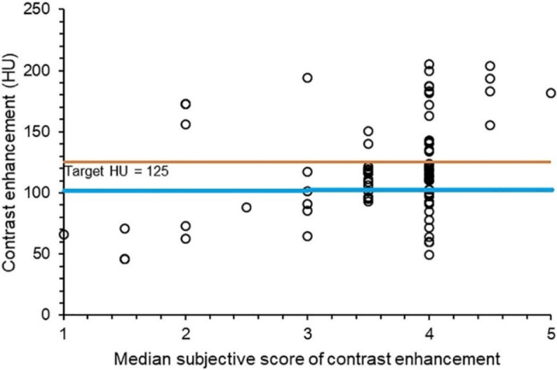
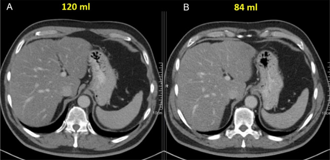
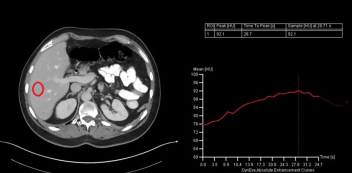
Objectives: to determine (1) a target Hounsfield unit (HU) for portal venous phase iodinated contrast media enhanced abdominal CT scans, (2) an equation for a personalized total contrast agent volume, and (3) the best/most appropriate time between injection and plateau/peak in HU enhancement. Material and methods: from an original dataset of 5,000 cases, a weight representative subset of 370 cases was sampled for detailed HU measurements. An additional 90 cases were used for visual grading to define the minimal HU required for diagnostic quality, which led to the proposed target HU. This study uses the fact that in a first approach, the injected contrast agent volume and HU correlate linearly. Based on the injected contrast agent volumes and HU measurements in the patient scans, it was then calculated which (ideal) volume would have reached the target value. The ideal volumes and patient data (weight, height, heart rate, age, and gender) were correlated by means of a regression analysis, to determine a new patient-specific contrast volume calculation equation. The best scan delay time was derived from the start of the injection to the HU enhancement plateau/peak evaluated from manually triggered venous phase scans. Results: The target HU value was 125. This can be achieved with a personalized contrast agent volume (ml), equal to - 108.5 + ∗ weight(kg) + 0.40 ∗ heart rate(bpm) + 0.61 ∗ height(cm). The time delay between injection and HU plateau/peak was found to be, on average, 102 s. Conclusion: this study proposes a comprehensive protocol for contrast enhanced venous phase scans, including a target HU, a personalized contrast volume, and a scan delay.
期刊介绍:
The purpose of the Journal of the Belgian Society of Radiology is the publication of articles dealing with diagnostic and interventional radiology, related imaging techniques, allied sciences, and continuing education.
 求助内容:
求助内容: 应助结果提醒方式:


