Yasmin Sadigh, Lailla Talbi, Juliette Monchen, Ayca Cozar, Kelsey Gori, Eelke M Bos, Ruben Dammers, Victor Volovici
{"title":"颅底训练外科医生行常规硬膜外视神经管减压术对前颅底脑膜瘤患者视力的影响。","authors":"Yasmin Sadigh, Lailla Talbi, Juliette Monchen, Ayca Cozar, Kelsey Gori, Eelke M Bos, Ruben Dammers, Victor Volovici","doi":"10.1007/s00701-025-06584-7","DOIUrl":null,"url":null,"abstract":"<p><strong>Purpose: </strong>Optic canal decompression is a surgical option in anterior skull base tumors with optic nerve involvement. Meningiomas may grow into the optic canal even without evidence of involvement on MRI studies. We aim to investigate the effect of routine optic canal unroofing performed by skull base trained surgeons versus general neurosurgeons on the postoperative visual outcomes in anterior skull base meningiomas.</p><p><strong>Methods: </strong>Between January 2013 and October 2023, consecutive patients in our institution who underwent craniotomies due to visual impairment were retrospectively reviewed. Patient records were screened for data on optic nerve compression, patient characteristics, lesion characteristics, intraoperative factors, the exact preoperative and postoperative visual acuity, as well as the postoperative clinical course. The primary outcome was the change in visual acuity postoperatively compared to the preoperative visual acuity. Multivariable linear regression analysis was performed with best postoperative visual acuity as a dependent adjusting for prognostic factors.</p><p><strong>Results: </strong>Out of 709 patients who underwent craniotomies for anterior skull base meningiomas, 94 patients showed optic nerve involvement on MRI. In total, 59 cases were treated by skull base trained surgeons and 35 by general neurosurgeons. Optic canal decompression was performed in 65% of the patients. There was no significant difference between patients treated by skull base surgeons and general neurosurgeons in terms of postoperative permanent complications. In patients with tuberculum sellae or anterior clinoid process meningiomas, postoperative secondary deterioration of visual acuity occurred in 40% (n = 10) of the cases treated by general neurosurgeons versus 11% (n = 4) in the group treated by skull base trained surgeons. In cases with a preoperative visual acuity of 0.2 or lower (35%, n = 33), 42% (n = 14) reached a best postoperative visual acuity of 0.5 or higher. Nineteen (20%) cases presented with functional blindness preoperatively. Of these, nine (47%) cases showed significant vision improvement postoperatively. Multivariable linear regression analysis revealed that patients with higher preoperative visual acuity reached a higher best visual acuity postoperatively.</p><p><strong>Conclusion: </strong>Patients with tuberculum sellae and anterior clinoid process meningiomas benefit from skull base surgeons trained in extradural optic canal decompression, as reflected by lower postoperative secondary visual acuity deterioration in patients treated by skull base trained surgeons. All cases presenting with tumors with optic apparatus involvement should be managed by skull base trained surgeons to maximize postoperative visual acuity preservation.</p>","PeriodicalId":7370,"journal":{"name":"Acta Neurochirurgica","volume":"167 1","pages":"170"},"PeriodicalIF":1.9000,"publicationDate":"2025-06-16","publicationTypes":"Journal Article","fieldsOfStudy":null,"isOpenAccess":false,"openAccessPdf":"https://www.ncbi.nlm.nih.gov/pmc/articles/PMC12170688/pdf/","citationCount":"0","resultStr":"{\"title\":\"Effect of routine extradural optic canal decompression performed by skull base trained surgeons on visual outcomes in patients with anterior skull base meningiomas.\",\"authors\":\"Yasmin Sadigh, Lailla Talbi, Juliette Monchen, Ayca Cozar, Kelsey Gori, Eelke M Bos, Ruben Dammers, Victor Volovici\",\"doi\":\"10.1007/s00701-025-06584-7\",\"DOIUrl\":null,\"url\":null,\"abstract\":\"<p><strong>Purpose: </strong>Optic canal decompression is a surgical option in anterior skull base tumors with optic nerve involvement. Meningiomas may grow into the optic canal even without evidence of involvement on MRI studies. We aim to investigate the effect of routine optic canal unroofing performed by skull base trained surgeons versus general neurosurgeons on the postoperative visual outcomes in anterior skull base meningiomas.</p><p><strong>Methods: </strong>Between January 2013 and October 2023, consecutive patients in our institution who underwent craniotomies due to visual impairment were retrospectively reviewed. Patient records were screened for data on optic nerve compression, patient characteristics, lesion characteristics, intraoperative factors, the exact preoperative and postoperative visual acuity, as well as the postoperative clinical course. The primary outcome was the change in visual acuity postoperatively compared to the preoperative visual acuity. Multivariable linear regression analysis was performed with best postoperative visual acuity as a dependent adjusting for prognostic factors.</p><p><strong>Results: </strong>Out of 709 patients who underwent craniotomies for anterior skull base meningiomas, 94 patients showed optic nerve involvement on MRI. In total, 59 cases were treated by skull base trained surgeons and 35 by general neurosurgeons. Optic canal decompression was performed in 65% of the patients. There was no significant difference between patients treated by skull base surgeons and general neurosurgeons in terms of postoperative permanent complications. In patients with tuberculum sellae or anterior clinoid process meningiomas, postoperative secondary deterioration of visual acuity occurred in 40% (n = 10) of the cases treated by general neurosurgeons versus 11% (n = 4) in the group treated by skull base trained surgeons. In cases with a preoperative visual acuity of 0.2 or lower (35%, n = 33), 42% (n = 14) reached a best postoperative visual acuity of 0.5 or higher. Nineteen (20%) cases presented with functional blindness preoperatively. Of these, nine (47%) cases showed significant vision improvement postoperatively. Multivariable linear regression analysis revealed that patients with higher preoperative visual acuity reached a higher best visual acuity postoperatively.</p><p><strong>Conclusion: </strong>Patients with tuberculum sellae and anterior clinoid process meningiomas benefit from skull base surgeons trained in extradural optic canal decompression, as reflected by lower postoperative secondary visual acuity deterioration in patients treated by skull base trained surgeons. All cases presenting with tumors with optic apparatus involvement should be managed by skull base trained surgeons to maximize postoperative visual acuity preservation.</p>\",\"PeriodicalId\":7370,\"journal\":{\"name\":\"Acta Neurochirurgica\",\"volume\":\"167 1\",\"pages\":\"170\"},\"PeriodicalIF\":1.9000,\"publicationDate\":\"2025-06-16\",\"publicationTypes\":\"Journal Article\",\"fieldsOfStudy\":null,\"isOpenAccess\":false,\"openAccessPdf\":\"https://www.ncbi.nlm.nih.gov/pmc/articles/PMC12170688/pdf/\",\"citationCount\":\"0\",\"resultStr\":null,\"platform\":\"Semanticscholar\",\"paperid\":null,\"PeriodicalName\":\"Acta Neurochirurgica\",\"FirstCategoryId\":\"3\",\"ListUrlMain\":\"https://doi.org/10.1007/s00701-025-06584-7\",\"RegionNum\":3,\"RegionCategory\":\"医学\",\"ArticlePicture\":[],\"TitleCN\":null,\"AbstractTextCN\":null,\"PMCID\":null,\"EPubDate\":\"\",\"PubModel\":\"\",\"JCR\":\"Q3\",\"JCRName\":\"CLINICAL NEUROLOGY\",\"Score\":null,\"Total\":0}","platform":"Semanticscholar","paperid":null,"PeriodicalName":"Acta Neurochirurgica","FirstCategoryId":"3","ListUrlMain":"https://doi.org/10.1007/s00701-025-06584-7","RegionNum":3,"RegionCategory":"医学","ArticlePicture":[],"TitleCN":null,"AbstractTextCN":null,"PMCID":null,"EPubDate":"","PubModel":"","JCR":"Q3","JCRName":"CLINICAL NEUROLOGY","Score":null,"Total":0}
Effect of routine extradural optic canal decompression performed by skull base trained surgeons on visual outcomes in patients with anterior skull base meningiomas.
Purpose: Optic canal decompression is a surgical option in anterior skull base tumors with optic nerve involvement. Meningiomas may grow into the optic canal even without evidence of involvement on MRI studies. We aim to investigate the effect of routine optic canal unroofing performed by skull base trained surgeons versus general neurosurgeons on the postoperative visual outcomes in anterior skull base meningiomas.
Methods: Between January 2013 and October 2023, consecutive patients in our institution who underwent craniotomies due to visual impairment were retrospectively reviewed. Patient records were screened for data on optic nerve compression, patient characteristics, lesion characteristics, intraoperative factors, the exact preoperative and postoperative visual acuity, as well as the postoperative clinical course. The primary outcome was the change in visual acuity postoperatively compared to the preoperative visual acuity. Multivariable linear regression analysis was performed with best postoperative visual acuity as a dependent adjusting for prognostic factors.
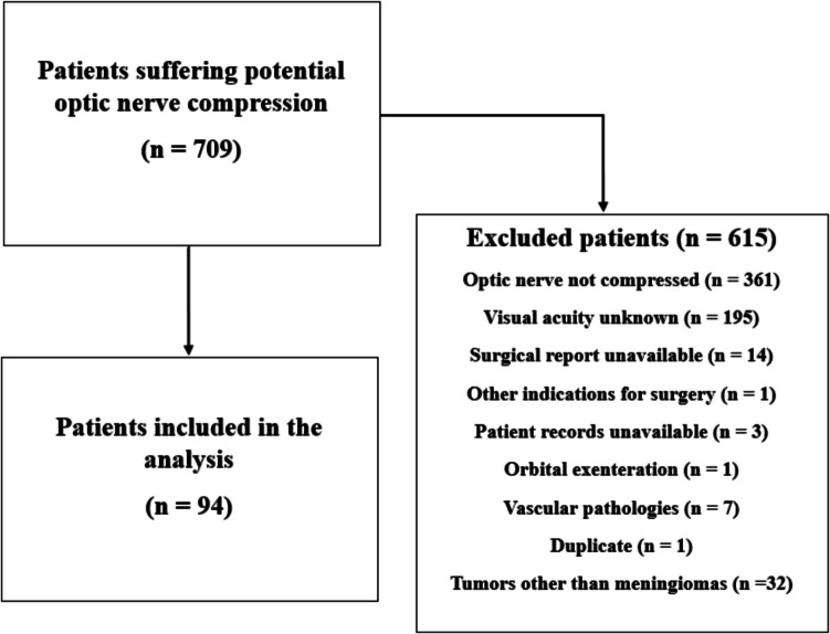
Results: Out of 709 patients who underwent craniotomies for anterior skull base meningiomas, 94 patients showed optic nerve involvement on MRI. In total, 59 cases were treated by skull base trained surgeons and 35 by general neurosurgeons. Optic canal decompression was performed in 65% of the patients. There was no significant difference between patients treated by skull base surgeons and general neurosurgeons in terms of postoperative permanent complications. In patients with tuberculum sellae or anterior clinoid process meningiomas, postoperative secondary deterioration of visual acuity occurred in 40% (n = 10) of the cases treated by general neurosurgeons versus 11% (n = 4) in the group treated by skull base trained surgeons. In cases with a preoperative visual acuity of 0.2 or lower (35%, n = 33), 42% (n = 14) reached a best postoperative visual acuity of 0.5 or higher. Nineteen (20%) cases presented with functional blindness preoperatively. Of these, nine (47%) cases showed significant vision improvement postoperatively. Multivariable linear regression analysis revealed that patients with higher preoperative visual acuity reached a higher best visual acuity postoperatively.
Conclusion: Patients with tuberculum sellae and anterior clinoid process meningiomas benefit from skull base surgeons trained in extradural optic canal decompression, as reflected by lower postoperative secondary visual acuity deterioration in patients treated by skull base trained surgeons. All cases presenting with tumors with optic apparatus involvement should be managed by skull base trained surgeons to maximize postoperative visual acuity preservation.
期刊介绍:
The journal "Acta Neurochirurgica" publishes only original papers useful both to research and clinical work. Papers should deal with clinical neurosurgery - diagnosis and diagnostic techniques, operative surgery and results, postoperative treatment - or with research work in neuroscience if the underlying questions or the results are of neurosurgical interest. Reports on congresses are given in brief accounts. As official organ of the European Association of Neurosurgical Societies the journal publishes all announcements of the E.A.N.S. and reports on the activities of its member societies. Only contributions written in English will be accepted.

 求助内容:
求助内容: 应助结果提醒方式:


