Luigi Tagliatesta, Matteo Arcari, Federico Guerri, Marco Lorenzoni, Jason M Jones
{"title":"锥形束CT三维重建在下颌阻生第三磨牙手术规划中的应用。","authors":"Luigi Tagliatesta, Matteo Arcari, Federico Guerri, Marco Lorenzoni, Jason M Jones","doi":"10.4103/njms.njms_135_22","DOIUrl":null,"url":null,"abstract":"<p><strong>Objectives: </strong>The aim of this work is to evaluate the possibility that the three-dimensional (3D) reconstruction of a cone beam computed tomography (CBCT) scan may facilitate the technical planning of the surgical extraction of third molars, whose roots are located in proximity of or in contact with the inferior alveolar nerve.</p><p><strong>Materials and methods: </strong>This article describes the planning process and the extraction of a lower third molar with the inferior alveolar nerve (IAN) entrapped through its roots using the 3D reconstruction of a CBCT scan.</p><p><strong>Results: </strong>After-surgery clinical examinations showed normal healing of the surgical wound and no neurological symptoms.</p><p><strong>Conclusions: </strong>The 3D reprocessing of a CBCT allows the construction of a computer-aided design model to investigate the spatial relationships between the wisdom tooth and the IAN. This method makes it possible to reconstruct a virtual model to preview the tooth and its spatial relationships, simplifying the mental processes of reworking and analyzing the existing relationships in the area, an important aid, especially in cases that are difficult to diagnose with a first-level examination. Indeed, it helps in reducing the risk of intra-operative injury to the nerve during extraction procedures.</p>","PeriodicalId":101444,"journal":{"name":"National journal of maxillofacial surgery","volume":"16 1","pages":"33-38"},"PeriodicalIF":0.0000,"publicationDate":"2025-01-01","publicationTypes":"Journal Article","fieldsOfStudy":null,"isOpenAccess":false,"openAccessPdf":"https://www.ncbi.nlm.nih.gov/pmc/articles/PMC12156845/pdf/","citationCount":"0","resultStr":"{\"title\":\"Three-dimensional reconstruction of cone beam CT scan in the planning of the surgery of mandibular impacted third molars.\",\"authors\":\"Luigi Tagliatesta, Matteo Arcari, Federico Guerri, Marco Lorenzoni, Jason M Jones\",\"doi\":\"10.4103/njms.njms_135_22\",\"DOIUrl\":null,\"url\":null,\"abstract\":\"<p><strong>Objectives: </strong>The aim of this work is to evaluate the possibility that the three-dimensional (3D) reconstruction of a cone beam computed tomography (CBCT) scan may facilitate the technical planning of the surgical extraction of third molars, whose roots are located in proximity of or in contact with the inferior alveolar nerve.</p><p><strong>Materials and methods: </strong>This article describes the planning process and the extraction of a lower third molar with the inferior alveolar nerve (IAN) entrapped through its roots using the 3D reconstruction of a CBCT scan.</p><p><strong>Results: </strong>After-surgery clinical examinations showed normal healing of the surgical wound and no neurological symptoms.</p><p><strong>Conclusions: </strong>The 3D reprocessing of a CBCT allows the construction of a computer-aided design model to investigate the spatial relationships between the wisdom tooth and the IAN. This method makes it possible to reconstruct a virtual model to preview the tooth and its spatial relationships, simplifying the mental processes of reworking and analyzing the existing relationships in the area, an important aid, especially in cases that are difficult to diagnose with a first-level examination. Indeed, it helps in reducing the risk of intra-operative injury to the nerve during extraction procedures.</p>\",\"PeriodicalId\":101444,\"journal\":{\"name\":\"National journal of maxillofacial surgery\",\"volume\":\"16 1\",\"pages\":\"33-38\"},\"PeriodicalIF\":0.0000,\"publicationDate\":\"2025-01-01\",\"publicationTypes\":\"Journal Article\",\"fieldsOfStudy\":null,\"isOpenAccess\":false,\"openAccessPdf\":\"https://www.ncbi.nlm.nih.gov/pmc/articles/PMC12156845/pdf/\",\"citationCount\":\"0\",\"resultStr\":null,\"platform\":\"Semanticscholar\",\"paperid\":null,\"PeriodicalName\":\"National journal of maxillofacial surgery\",\"FirstCategoryId\":\"1085\",\"ListUrlMain\":\"https://doi.org/10.4103/njms.njms_135_22\",\"RegionNum\":0,\"RegionCategory\":null,\"ArticlePicture\":[],\"TitleCN\":null,\"AbstractTextCN\":null,\"PMCID\":null,\"EPubDate\":\"2025/4/28 0:00:00\",\"PubModel\":\"Epub\",\"JCR\":\"\",\"JCRName\":\"\",\"Score\":null,\"Total\":0}","platform":"Semanticscholar","paperid":null,"PeriodicalName":"National journal of maxillofacial surgery","FirstCategoryId":"1085","ListUrlMain":"https://doi.org/10.4103/njms.njms_135_22","RegionNum":0,"RegionCategory":null,"ArticlePicture":[],"TitleCN":null,"AbstractTextCN":null,"PMCID":null,"EPubDate":"2025/4/28 0:00:00","PubModel":"Epub","JCR":"","JCRName":"","Score":null,"Total":0}
Three-dimensional reconstruction of cone beam CT scan in the planning of the surgery of mandibular impacted third molars.
Objectives: The aim of this work is to evaluate the possibility that the three-dimensional (3D) reconstruction of a cone beam computed tomography (CBCT) scan may facilitate the technical planning of the surgical extraction of third molars, whose roots are located in proximity of or in contact with the inferior alveolar nerve.
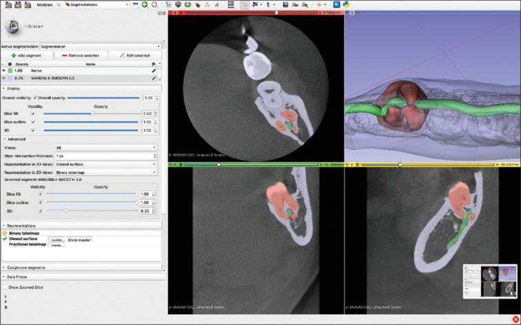
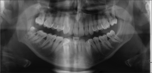
Materials and methods: This article describes the planning process and the extraction of a lower third molar with the inferior alveolar nerve (IAN) entrapped through its roots using the 3D reconstruction of a CBCT scan.
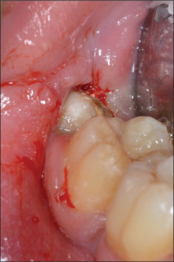
Results: After-surgery clinical examinations showed normal healing of the surgical wound and no neurological symptoms.
Conclusions: The 3D reprocessing of a CBCT allows the construction of a computer-aided design model to investigate the spatial relationships between the wisdom tooth and the IAN. This method makes it possible to reconstruct a virtual model to preview the tooth and its spatial relationships, simplifying the mental processes of reworking and analyzing the existing relationships in the area, an important aid, especially in cases that are difficult to diagnose with a first-level examination. Indeed, it helps in reducing the risk of intra-operative injury to the nerve during extraction procedures.

 求助内容:
求助内容: 应助结果提醒方式:


