Thomas P A Baltes, Feriel Dalansi, Maryam R Al-Naimi, Marcelo Bordalo, Louis Holtzhausen, Rod Whiteley, Marco Cardinale, Pieter D'Hooghe, Gino M M J Kerkhoffs, Johannes L Tol
{"title":"急性踝关节韧带损伤运动员软骨和骨软骨病变的发生率、大小和解剖位置。","authors":"Thomas P A Baltes, Feriel Dalansi, Maryam R Al-Naimi, Marcelo Bordalo, Louis Holtzhausen, Rod Whiteley, Marco Cardinale, Pieter D'Hooghe, Gino M M J Kerkhoffs, Johannes L Tol","doi":"10.1177/03635465251344187","DOIUrl":null,"url":null,"abstract":"<p><strong>Background: </strong>In athletes with an acute ligamentous ankle injury, cartilage and osteochondral lesions ([O]CLs) have been reported in 8% using 1.5-T magnetic resonance imaging (MRI). Visualization of cartilage injuries improves with the use of higher field strengths.</p><p><strong>Purpose: </strong>To evaluate the prevalence, size, and anatomic location of (O)CLs in athletes with an acute ligamentous ankle injury using 3-T MRI, as well as to determine the association of (O)CLs with injury of (1) the lateral ankle ligaments and (2) anterior syndesmosis.</p><p><strong>Study design: </strong>Cohort study; Level of evidence, 3.</p><p><strong>Methods: </strong>For this prospective cohort study, all acute ligamentous ankle injuries in athletes (≥18 years of age) evaluated in the outpatient department of a specialized orthopaedic and sports medicine hospital within 7 days after injury were assed for eligibility. Acute ankle injuries were excluded if 3-T MRI could not be obtained within 10 days after injury or if imaging demonstrated a frank fracture. A musculoskeletal radiologist assessed MRI scans for the presence, location, and size of (O)CLs. Morphology was graded using the modified Berndt and Harty score, Griffith MRI score, and International Cartilage Regeneration & Joint Preservation Society score. In addition, injuries of the lateral ankle ligaments and anterior syndesmosis were graded. A multivariate logistic regression analysis was performed to evaluate the association between (O)CLs and injury of the (1) lateral ankle ligaments and (2) anterior syndesmosis.</p><p><strong>Results: </strong>Between September 2016 and February 2020, 171 acute ankle injuries in 166 athletes were included in this study. The overall prevalence of (O)CLs was 14%. (O)CLs of the talus and tibia were observed in 24 (14%) and 9 (5%) acute ankle injuries, respectively. Of 33 (O)CLs, 28 (85%) were classified as cartilage lesions. Lateral ligament injury was observed in 73% of acute ankle injuries, and anterior syndesmosis injury in 38%. Multivariate logistic regression analysis did not show significantly higher odds of (O)CLs in the presence of anterior syndesmosis injury (OR, 2.16; 95% CI, 0.90-5.16).</p><p><strong>Conclusion: </strong>In athletes with an acute ligamentous ankle injury, a prevalence for (O)CLs of 14% was established using 3-T MRI. The majority were cartilage lesions. No statistically significant association was established between (O)CLs and lateral ligament or syndesmosis injury was established.</p>","PeriodicalId":55528,"journal":{"name":"American Journal of Sports Medicine","volume":" ","pages":"2173-2180"},"PeriodicalIF":4.5000,"publicationDate":"2025-07-01","publicationTypes":"Journal Article","fieldsOfStudy":null,"isOpenAccess":false,"openAccessPdf":"https://www.ncbi.nlm.nih.gov/pmc/articles/PMC12235055/pdf/","citationCount":"0","resultStr":"{\"title\":\"The Prevalence, Size, and Anatomic Location of Cartilage and Osteochondral Lesions in Athletes With an Acute Ligamentous Ankle Injury.\",\"authors\":\"Thomas P A Baltes, Feriel Dalansi, Maryam R Al-Naimi, Marcelo Bordalo, Louis Holtzhausen, Rod Whiteley, Marco Cardinale, Pieter D'Hooghe, Gino M M J Kerkhoffs, Johannes L Tol\",\"doi\":\"10.1177/03635465251344187\",\"DOIUrl\":null,\"url\":null,\"abstract\":\"<p><strong>Background: </strong>In athletes with an acute ligamentous ankle injury, cartilage and osteochondral lesions ([O]CLs) have been reported in 8% using 1.5-T magnetic resonance imaging (MRI). Visualization of cartilage injuries improves with the use of higher field strengths.</p><p><strong>Purpose: </strong>To evaluate the prevalence, size, and anatomic location of (O)CLs in athletes with an acute ligamentous ankle injury using 3-T MRI, as well as to determine the association of (O)CLs with injury of (1) the lateral ankle ligaments and (2) anterior syndesmosis.</p><p><strong>Study design: </strong>Cohort study; Level of evidence, 3.</p><p><strong>Methods: </strong>For this prospective cohort study, all acute ligamentous ankle injuries in athletes (≥18 years of age) evaluated in the outpatient department of a specialized orthopaedic and sports medicine hospital within 7 days after injury were assed for eligibility. Acute ankle injuries were excluded if 3-T MRI could not be obtained within 10 days after injury or if imaging demonstrated a frank fracture. A musculoskeletal radiologist assessed MRI scans for the presence, location, and size of (O)CLs. Morphology was graded using the modified Berndt and Harty score, Griffith MRI score, and International Cartilage Regeneration & Joint Preservation Society score. In addition, injuries of the lateral ankle ligaments and anterior syndesmosis were graded. A multivariate logistic regression analysis was performed to evaluate the association between (O)CLs and injury of the (1) lateral ankle ligaments and (2) anterior syndesmosis.</p><p><strong>Results: </strong>Between September 2016 and February 2020, 171 acute ankle injuries in 166 athletes were included in this study. The overall prevalence of (O)CLs was 14%. (O)CLs of the talus and tibia were observed in 24 (14%) and 9 (5%) acute ankle injuries, respectively. Of 33 (O)CLs, 28 (85%) were classified as cartilage lesions. Lateral ligament injury was observed in 73% of acute ankle injuries, and anterior syndesmosis injury in 38%. Multivariate logistic regression analysis did not show significantly higher odds of (O)CLs in the presence of anterior syndesmosis injury (OR, 2.16; 95% CI, 0.90-5.16).</p><p><strong>Conclusion: </strong>In athletes with an acute ligamentous ankle injury, a prevalence for (O)CLs of 14% was established using 3-T MRI. The majority were cartilage lesions. No statistically significant association was established between (O)CLs and lateral ligament or syndesmosis injury was established.</p>\",\"PeriodicalId\":55528,\"journal\":{\"name\":\"American Journal of Sports Medicine\",\"volume\":\" \",\"pages\":\"2173-2180\"},\"PeriodicalIF\":4.5000,\"publicationDate\":\"2025-07-01\",\"publicationTypes\":\"Journal Article\",\"fieldsOfStudy\":null,\"isOpenAccess\":false,\"openAccessPdf\":\"https://www.ncbi.nlm.nih.gov/pmc/articles/PMC12235055/pdf/\",\"citationCount\":\"0\",\"resultStr\":null,\"platform\":\"Semanticscholar\",\"paperid\":null,\"PeriodicalName\":\"American Journal of Sports Medicine\",\"FirstCategoryId\":\"3\",\"ListUrlMain\":\"https://doi.org/10.1177/03635465251344187\",\"RegionNum\":1,\"RegionCategory\":\"医学\",\"ArticlePicture\":[],\"TitleCN\":null,\"AbstractTextCN\":null,\"PMCID\":null,\"EPubDate\":\"2025/6/12 0:00:00\",\"PubModel\":\"Epub\",\"JCR\":\"Q1\",\"JCRName\":\"ORTHOPEDICS\",\"Score\":null,\"Total\":0}","platform":"Semanticscholar","paperid":null,"PeriodicalName":"American Journal of Sports Medicine","FirstCategoryId":"3","ListUrlMain":"https://doi.org/10.1177/03635465251344187","RegionNum":1,"RegionCategory":"医学","ArticlePicture":[],"TitleCN":null,"AbstractTextCN":null,"PMCID":null,"EPubDate":"2025/6/12 0:00:00","PubModel":"Epub","JCR":"Q1","JCRName":"ORTHOPEDICS","Score":null,"Total":0}
The Prevalence, Size, and Anatomic Location of Cartilage and Osteochondral Lesions in Athletes With an Acute Ligamentous Ankle Injury.
Background: In athletes with an acute ligamentous ankle injury, cartilage and osteochondral lesions ([O]CLs) have been reported in 8% using 1.5-T magnetic resonance imaging (MRI). Visualization of cartilage injuries improves with the use of higher field strengths.
Purpose: To evaluate the prevalence, size, and anatomic location of (O)CLs in athletes with an acute ligamentous ankle injury using 3-T MRI, as well as to determine the association of (O)CLs with injury of (1) the lateral ankle ligaments and (2) anterior syndesmosis.
Study design: Cohort study; Level of evidence, 3.
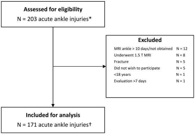
Methods: For this prospective cohort study, all acute ligamentous ankle injuries in athletes (≥18 years of age) evaluated in the outpatient department of a specialized orthopaedic and sports medicine hospital within 7 days after injury were assed for eligibility. Acute ankle injuries were excluded if 3-T MRI could not be obtained within 10 days after injury or if imaging demonstrated a frank fracture. A musculoskeletal radiologist assessed MRI scans for the presence, location, and size of (O)CLs. Morphology was graded using the modified Berndt and Harty score, Griffith MRI score, and International Cartilage Regeneration & Joint Preservation Society score. In addition, injuries of the lateral ankle ligaments and anterior syndesmosis were graded. A multivariate logistic regression analysis was performed to evaluate the association between (O)CLs and injury of the (1) lateral ankle ligaments and (2) anterior syndesmosis.
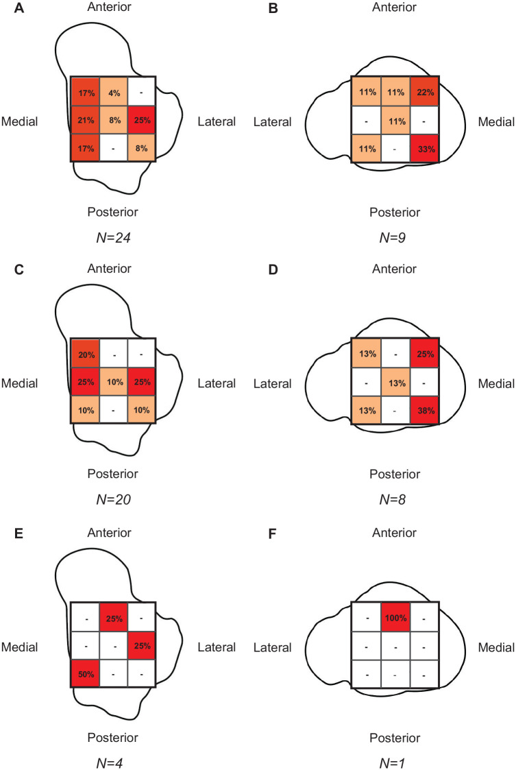
Results: Between September 2016 and February 2020, 171 acute ankle injuries in 166 athletes were included in this study. The overall prevalence of (O)CLs was 14%. (O)CLs of the talus and tibia were observed in 24 (14%) and 9 (5%) acute ankle injuries, respectively. Of 33 (O)CLs, 28 (85%) were classified as cartilage lesions. Lateral ligament injury was observed in 73% of acute ankle injuries, and anterior syndesmosis injury in 38%. Multivariate logistic regression analysis did not show significantly higher odds of (O)CLs in the presence of anterior syndesmosis injury (OR, 2.16; 95% CI, 0.90-5.16).
Conclusion: In athletes with an acute ligamentous ankle injury, a prevalence for (O)CLs of 14% was established using 3-T MRI. The majority were cartilage lesions. No statistically significant association was established between (O)CLs and lateral ligament or syndesmosis injury was established.
期刊介绍:
An invaluable resource for the orthopaedic sports medicine community, _The American Journal of Sports Medicine_ is a peer-reviewed scientific journal, first published in 1972. It is the official publication of the [American Orthopaedic Society for Sports Medicine (AOSSM)](http://www.sportsmed.org/)! The journal acts as an important forum for independent orthopaedic sports medicine research and education, allowing clinical practitioners the ability to make decisions based on sound scientific information.
This journal is a must-read for:
* Orthopaedic Surgeons and Specialists
* Sports Medicine Physicians
* Physiatrists
* Athletic Trainers
* Team Physicians
* And Physical Therapists


 求助内容:
求助内容: 应助结果提醒方式:


