{"title":"颈椎椎弓根螺钉置入的术前规划:识别关键形态学参数。","authors":"Yuya Okada, Hiroaki Nakashima, Sadayuki Ito, Naoki Segi, Jun Ouchida, Tetsuya Urasaki, Shiro Imagama","doi":"10.22603/ssrr.2024-0243","DOIUrl":null,"url":null,"abstract":"<p><strong>Introduction: </strong>Cervical pedicle screw (CPS) placement is crucial for posterior cervical fusion surgery due to its strong fixation ability. However, CPS insertion is associated with risks, including screw perforation, which can lead to complications such as vertebral artery injury and neurological deficits. Although previous studies have explored some morphological factors affecting CPS placement, comprehensive data on specific parameters contributing to perforation remains limited. This study aimed to investigate cervical vertebrae features associated with CPS perforation and established threshold values for improved preoperative planning.</p><p><strong>Methods: </strong>A retrospective analysis of 36 patients who underwent posterior cervical fusion surgery with CPS placement was conducted using preoperative computed tomography (CT)-based navigation. Cases with CPS insertion at C1 or C2 were excluded. The key morphological parameters-optimal screw trajectory angle, pedicle diameter, and distance from the entry point to the pedicle isthmus (DEP)-were measured on preoperative CT images. CPS placement accuracy was assessed postoperatively using Neo's classification. The receiver operating characteristic (ROC) curve analysis determined the cutoff values for predicting CPS perforation.</p><p><strong>Results: </strong>Among the 102 CPSs placed from C3 to C7, the overall perforation rate was 25.5%. C3 had the highest perforation rate (45.5%), whereas C7 had the lowest (3.1%). The vertebrae with CPS perforation exhibited a significantly larger optimal screw trajectory angle (45.5° vs. 38.0°, p<0.001), smaller pedicle diameter (4.2 mm vs. 5.2 mm, p<0.001), and longer DEP (13.2 mm vs. 11.9 mm, p=0.002). The ROC analysis identified the following cutoff values: 44.0° for the optimal angle, 4.35 mm for the pedicle diameter, and 12.7 mm for the DEP. These morphological parameters strongly predicted the risk of CPS perforation.</p><p><strong>Conclusions: </strong>Establishing key morphological thresholds enhances preoperative planning for CPS placement, improves accuracy and patient safety, and minimizes complications.</p>","PeriodicalId":22253,"journal":{"name":"Spine Surgery and Related Research","volume":"9 3","pages":"313-320"},"PeriodicalIF":1.2000,"publicationDate":"2024-12-20","publicationTypes":"Journal Article","fieldsOfStudy":null,"isOpenAccess":false,"openAccessPdf":"https://www.ncbi.nlm.nih.gov/pmc/articles/PMC12151281/pdf/","citationCount":"0","resultStr":"{\"title\":\"Preoperative Planning for Cervical Pedicle Screw Placement: Identifying Key Morphological Parameters.\",\"authors\":\"Yuya Okada, Hiroaki Nakashima, Sadayuki Ito, Naoki Segi, Jun Ouchida, Tetsuya Urasaki, Shiro Imagama\",\"doi\":\"10.22603/ssrr.2024-0243\",\"DOIUrl\":null,\"url\":null,\"abstract\":\"<p><strong>Introduction: </strong>Cervical pedicle screw (CPS) placement is crucial for posterior cervical fusion surgery due to its strong fixation ability. However, CPS insertion is associated with risks, including screw perforation, which can lead to complications such as vertebral artery injury and neurological deficits. Although previous studies have explored some morphological factors affecting CPS placement, comprehensive data on specific parameters contributing to perforation remains limited. This study aimed to investigate cervical vertebrae features associated with CPS perforation and established threshold values for improved preoperative planning.</p><p><strong>Methods: </strong>A retrospective analysis of 36 patients who underwent posterior cervical fusion surgery with CPS placement was conducted using preoperative computed tomography (CT)-based navigation. Cases with CPS insertion at C1 or C2 were excluded. The key morphological parameters-optimal screw trajectory angle, pedicle diameter, and distance from the entry point to the pedicle isthmus (DEP)-were measured on preoperative CT images. CPS placement accuracy was assessed postoperatively using Neo's classification. The receiver operating characteristic (ROC) curve analysis determined the cutoff values for predicting CPS perforation.</p><p><strong>Results: </strong>Among the 102 CPSs placed from C3 to C7, the overall perforation rate was 25.5%. C3 had the highest perforation rate (45.5%), whereas C7 had the lowest (3.1%). The vertebrae with CPS perforation exhibited a significantly larger optimal screw trajectory angle (45.5° vs. 38.0°, p<0.001), smaller pedicle diameter (4.2 mm vs. 5.2 mm, p<0.001), and longer DEP (13.2 mm vs. 11.9 mm, p=0.002). The ROC analysis identified the following cutoff values: 44.0° for the optimal angle, 4.35 mm for the pedicle diameter, and 12.7 mm for the DEP. These morphological parameters strongly predicted the risk of CPS perforation.</p><p><strong>Conclusions: </strong>Establishing key morphological thresholds enhances preoperative planning for CPS placement, improves accuracy and patient safety, and minimizes complications.</p>\",\"PeriodicalId\":22253,\"journal\":{\"name\":\"Spine Surgery and Related Research\",\"volume\":\"9 3\",\"pages\":\"313-320\"},\"PeriodicalIF\":1.2000,\"publicationDate\":\"2024-12-20\",\"publicationTypes\":\"Journal Article\",\"fieldsOfStudy\":null,\"isOpenAccess\":false,\"openAccessPdf\":\"https://www.ncbi.nlm.nih.gov/pmc/articles/PMC12151281/pdf/\",\"citationCount\":\"0\",\"resultStr\":null,\"platform\":\"Semanticscholar\",\"paperid\":null,\"PeriodicalName\":\"Spine Surgery and Related Research\",\"FirstCategoryId\":\"1085\",\"ListUrlMain\":\"https://doi.org/10.22603/ssrr.2024-0243\",\"RegionNum\":0,\"RegionCategory\":null,\"ArticlePicture\":[],\"TitleCN\":null,\"AbstractTextCN\":null,\"PMCID\":null,\"EPubDate\":\"2025/5/27 0:00:00\",\"PubModel\":\"eCollection\",\"JCR\":\"Q3\",\"JCRName\":\"SURGERY\",\"Score\":null,\"Total\":0}","platform":"Semanticscholar","paperid":null,"PeriodicalName":"Spine Surgery and Related Research","FirstCategoryId":"1085","ListUrlMain":"https://doi.org/10.22603/ssrr.2024-0243","RegionNum":0,"RegionCategory":null,"ArticlePicture":[],"TitleCN":null,"AbstractTextCN":null,"PMCID":null,"EPubDate":"2025/5/27 0:00:00","PubModel":"eCollection","JCR":"Q3","JCRName":"SURGERY","Score":null,"Total":0}
Introduction: Cervical pedicle screw (CPS) placement is crucial for posterior cervical fusion surgery due to its strong fixation ability. However, CPS insertion is associated with risks, including screw perforation, which can lead to complications such as vertebral artery injury and neurological deficits. Although previous studies have explored some morphological factors affecting CPS placement, comprehensive data on specific parameters contributing to perforation remains limited. This study aimed to investigate cervical vertebrae features associated with CPS perforation and established threshold values for improved preoperative planning.
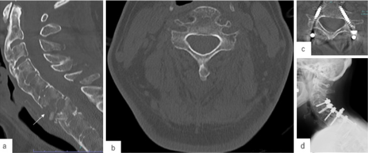
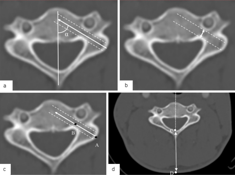
Methods: A retrospective analysis of 36 patients who underwent posterior cervical fusion surgery with CPS placement was conducted using preoperative computed tomography (CT)-based navigation. Cases with CPS insertion at C1 or C2 were excluded. The key morphological parameters-optimal screw trajectory angle, pedicle diameter, and distance from the entry point to the pedicle isthmus (DEP)-were measured on preoperative CT images. CPS placement accuracy was assessed postoperatively using Neo's classification. The receiver operating characteristic (ROC) curve analysis determined the cutoff values for predicting CPS perforation.
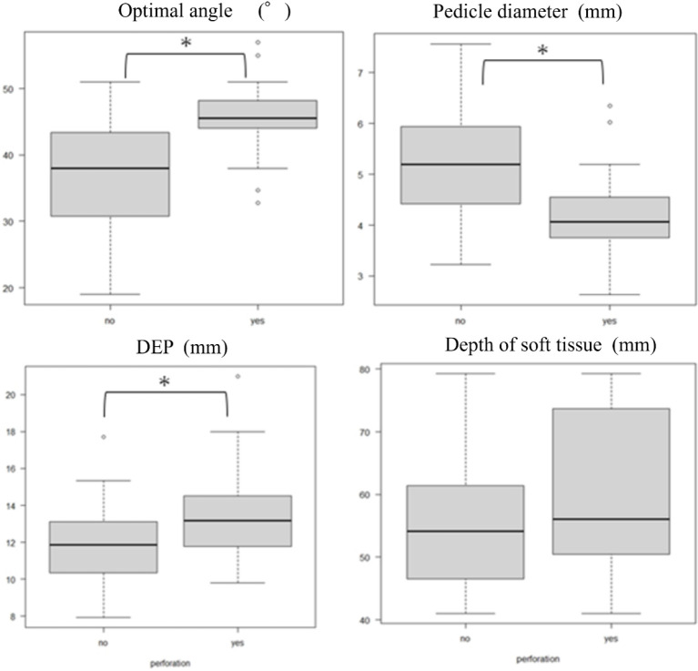
Results: Among the 102 CPSs placed from C3 to C7, the overall perforation rate was 25.5%. C3 had the highest perforation rate (45.5%), whereas C7 had the lowest (3.1%). The vertebrae with CPS perforation exhibited a significantly larger optimal screw trajectory angle (45.5° vs. 38.0°, p<0.001), smaller pedicle diameter (4.2 mm vs. 5.2 mm, p<0.001), and longer DEP (13.2 mm vs. 11.9 mm, p=0.002). The ROC analysis identified the following cutoff values: 44.0° for the optimal angle, 4.35 mm for the pedicle diameter, and 12.7 mm for the DEP. These morphological parameters strongly predicted the risk of CPS perforation.
Conclusions: Establishing key morphological thresholds enhances preoperative planning for CPS placement, improves accuracy and patient safety, and minimizes complications.

 求助内容:
求助内容: 应助结果提醒方式:


