Julia Mestre, Lorea Chaparro-González, Isabel Granada, Mar Mallo, Emili Cid, Estefania Mancini, Oriol Calvete, Ruth M Risueño, Daniel T Starczynowski, Francesc Solé
{"title":"MDS-L细胞系的综合细胞遗传学和基因组谱分析。","authors":"Julia Mestre, Lorea Chaparro-González, Isabel Granada, Mar Mallo, Emili Cid, Estefania Mancini, Oriol Calvete, Ruth M Risueño, Daniel T Starczynowski, Francesc Solé","doi":"10.1186/s13039-025-00714-7","DOIUrl":null,"url":null,"abstract":"<p><p>Among the human leukemia cell lines described in the literature, only the MDS-L cell line has been definitively established from a patient during the myelodysplastic syndrome (MDS) phase of the disease. However, the limited studies on its genomic complexity have restricted its applicability as an in vitro model for MDS. Here, we aimed to better characterize the chromosomal and genetic alterations of MDS-L. A comprehensive approach was employed combining conventional G banding, multicolor FISH (M-FISH), SNP arrays with the novel Optical Genome Mapping (OGM) technology. In addition, the mutational landscape was defined using targeted next-generation sequencing (NGS). G-banding revealed two karyotypically distinct cell populations, both exhibiting complex karyotypes. Using G-banding and OGM, we identified previously undescribed structural alterations, including der(1)t(1;7)(q11;q11.2), del(1)(q11), der(4)t(4;5)(p16;q11.2), i(5)(p10), der(6)t(6;15)(p21.3;q15), i(8)(q10), der(9)t(9;10)(q34;p11.21), der(19)t(6;19)(p13;p22) and i(22)(q10). Both OGM and SNP microarray analyses detected multiple copy number variants and regions of homozygosity. Chromosome breakpoints were precisely defined by OGM, allowing the identification of gene disruption events. Moreover, M-FISH technique validated the origins of additional chromosomal material observed in the karyotype, identified cryptic rearrangements, and distinguished the two clonal populations within the cell line. Finally, NGS revealed mutations in CEBPA, NRAS, TET2 and TP53 genes associated with MDS pathology. This multi-technique approach has enabled a precise characterization of the MDS-L cell line's genomic complexity, highlighting the unique contributions of each technique in uncovering various genetic alterations and establishing a valuable resource for mechanistic studies and pre-clinical drug development.</p>","PeriodicalId":19099,"journal":{"name":"Molecular Cytogenetics","volume":"18 1","pages":"11"},"PeriodicalIF":1.4000,"publicationDate":"2025-06-11","publicationTypes":"Journal Article","fieldsOfStudy":null,"isOpenAccess":false,"openAccessPdf":"https://www.ncbi.nlm.nih.gov/pmc/articles/PMC12160373/pdf/","citationCount":"0","resultStr":"{\"title\":\"Integrated cytogenetic and genomic profiling of the MDS-L cell line.\",\"authors\":\"Julia Mestre, Lorea Chaparro-González, Isabel Granada, Mar Mallo, Emili Cid, Estefania Mancini, Oriol Calvete, Ruth M Risueño, Daniel T Starczynowski, Francesc Solé\",\"doi\":\"10.1186/s13039-025-00714-7\",\"DOIUrl\":null,\"url\":null,\"abstract\":\"<p><p>Among the human leukemia cell lines described in the literature, only the MDS-L cell line has been definitively established from a patient during the myelodysplastic syndrome (MDS) phase of the disease. However, the limited studies on its genomic complexity have restricted its applicability as an in vitro model for MDS. Here, we aimed to better characterize the chromosomal and genetic alterations of MDS-L. A comprehensive approach was employed combining conventional G banding, multicolor FISH (M-FISH), SNP arrays with the novel Optical Genome Mapping (OGM) technology. In addition, the mutational landscape was defined using targeted next-generation sequencing (NGS). G-banding revealed two karyotypically distinct cell populations, both exhibiting complex karyotypes. Using G-banding and OGM, we identified previously undescribed structural alterations, including der(1)t(1;7)(q11;q11.2), del(1)(q11), der(4)t(4;5)(p16;q11.2), i(5)(p10), der(6)t(6;15)(p21.3;q15), i(8)(q10), der(9)t(9;10)(q34;p11.21), der(19)t(6;19)(p13;p22) and i(22)(q10). Both OGM and SNP microarray analyses detected multiple copy number variants and regions of homozygosity. Chromosome breakpoints were precisely defined by OGM, allowing the identification of gene disruption events. Moreover, M-FISH technique validated the origins of additional chromosomal material observed in the karyotype, identified cryptic rearrangements, and distinguished the two clonal populations within the cell line. Finally, NGS revealed mutations in CEBPA, NRAS, TET2 and TP53 genes associated with MDS pathology. This multi-technique approach has enabled a precise characterization of the MDS-L cell line's genomic complexity, highlighting the unique contributions of each technique in uncovering various genetic alterations and establishing a valuable resource for mechanistic studies and pre-clinical drug development.</p>\",\"PeriodicalId\":19099,\"journal\":{\"name\":\"Molecular Cytogenetics\",\"volume\":\"18 1\",\"pages\":\"11\"},\"PeriodicalIF\":1.4000,\"publicationDate\":\"2025-06-11\",\"publicationTypes\":\"Journal Article\",\"fieldsOfStudy\":null,\"isOpenAccess\":false,\"openAccessPdf\":\"https://www.ncbi.nlm.nih.gov/pmc/articles/PMC12160373/pdf/\",\"citationCount\":\"0\",\"resultStr\":null,\"platform\":\"Semanticscholar\",\"paperid\":null,\"PeriodicalName\":\"Molecular Cytogenetics\",\"FirstCategoryId\":\"99\",\"ListUrlMain\":\"https://doi.org/10.1186/s13039-025-00714-7\",\"RegionNum\":4,\"RegionCategory\":\"生物学\",\"ArticlePicture\":[],\"TitleCN\":null,\"AbstractTextCN\":null,\"PMCID\":null,\"EPubDate\":\"\",\"PubModel\":\"\",\"JCR\":\"Q4\",\"JCRName\":\"GENETICS & HEREDITY\",\"Score\":null,\"Total\":0}","platform":"Semanticscholar","paperid":null,"PeriodicalName":"Molecular Cytogenetics","FirstCategoryId":"99","ListUrlMain":"https://doi.org/10.1186/s13039-025-00714-7","RegionNum":4,"RegionCategory":"生物学","ArticlePicture":[],"TitleCN":null,"AbstractTextCN":null,"PMCID":null,"EPubDate":"","PubModel":"","JCR":"Q4","JCRName":"GENETICS & HEREDITY","Score":null,"Total":0}
Integrated cytogenetic and genomic profiling of the MDS-L cell line.
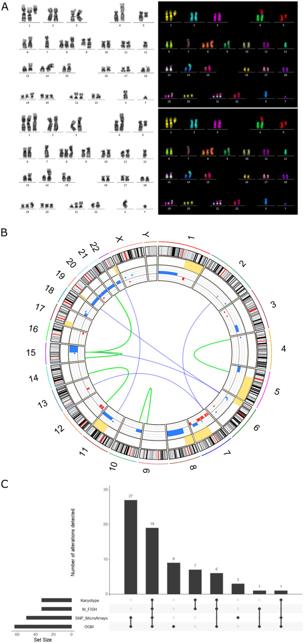
Among the human leukemia cell lines described in the literature, only the MDS-L cell line has been definitively established from a patient during the myelodysplastic syndrome (MDS) phase of the disease. However, the limited studies on its genomic complexity have restricted its applicability as an in vitro model for MDS. Here, we aimed to better characterize the chromosomal and genetic alterations of MDS-L. A comprehensive approach was employed combining conventional G banding, multicolor FISH (M-FISH), SNP arrays with the novel Optical Genome Mapping (OGM) technology. In addition, the mutational landscape was defined using targeted next-generation sequencing (NGS). G-banding revealed two karyotypically distinct cell populations, both exhibiting complex karyotypes. Using G-banding and OGM, we identified previously undescribed structural alterations, including der(1)t(1;7)(q11;q11.2), del(1)(q11), der(4)t(4;5)(p16;q11.2), i(5)(p10), der(6)t(6;15)(p21.3;q15), i(8)(q10), der(9)t(9;10)(q34;p11.21), der(19)t(6;19)(p13;p22) and i(22)(q10). Both OGM and SNP microarray analyses detected multiple copy number variants and regions of homozygosity. Chromosome breakpoints were precisely defined by OGM, allowing the identification of gene disruption events. Moreover, M-FISH technique validated the origins of additional chromosomal material observed in the karyotype, identified cryptic rearrangements, and distinguished the two clonal populations within the cell line. Finally, NGS revealed mutations in CEBPA, NRAS, TET2 and TP53 genes associated with MDS pathology. This multi-technique approach has enabled a precise characterization of the MDS-L cell line's genomic complexity, highlighting the unique contributions of each technique in uncovering various genetic alterations and establishing a valuable resource for mechanistic studies and pre-clinical drug development.
期刊介绍:
Molecular Cytogenetics encompasses all aspects of chromosome biology and the application of molecular cytogenetic techniques in all areas of biology and medicine, including structural and functional organization of the chromosome and nucleus, genome variation, expression and evolution, chromosome abnormalities and genomic variations in medical genetics and tumor genetics.
Molecular Cytogenetics primarily defines a large set of the techniques that operate either with the entire genome or with specific targeted DNA sequences. Topical areas include, but are not limited to:
-Structural and functional organization of chromosome and nucleus-
Genome variation, expression and evolution-
Animal and plant molecular cytogenetics and genomics-
Chromosome abnormalities and genomic variations in clinical genetics-
Applications in preimplantation, pre- and post-natal diagnosis-
Applications in the central nervous system, cancer and haematology research-
Previously unreported applications of molecular cytogenetic techniques-
Development of new techniques or significant enhancements to established techniques.
This journal is a source for numerous scientists all over the world, who wish to improve or introduce molecular cytogenetic techniques into their practice.
 求助内容:
求助内容: 应助结果提醒方式:


