Thorbjørn Erik Køppen Christensen, Takeshi Moriishi and Toshihisa Komori
{"title":"羟基磷灰石晶体在整个小鼠股骨上的定向成像。","authors":"Thorbjørn Erik Køppen Christensen, Takeshi Moriishi and Toshihisa Komori","doi":"10.1039/D5FD00009B","DOIUrl":null,"url":null,"abstract":"<p >Imaging the orientations of crystallites in bone requires the usage of synchrotron X-ray radiation, which is a limited resource for researchers. Thus scans have historically been limited to either small regions or few samples. In the present study, we scan 16 full frontal cross sections of mouse femora. This makes it possible to study structure, orientation, and composition, statistically across many different bones and animals, while preserving the structural context. From the following analysis, we can deduce that while the trabecular bone in the shaft has a larger fraction of oriented crystallites than other regions in the bone, the oriented fraction is more well aligned in the cortical bone in the shaft compared to other regions in the bone. We also see that the crystallites in the cortical and trabecular bone are longer than those in the femoral head and the condyle. The study also shows a larger Sr content in the cortical bone compared to other regions, and a larger Zn content in the femoral head compared to other regions of the bones. This study shows the need for and possibility of scanning larger regions to understand bioinorganic materials.</p>","PeriodicalId":49075,"journal":{"name":"Faraday Discussions","volume":"261 ","pages":" 446-460"},"PeriodicalIF":3.1000,"publicationDate":"2025-04-04","publicationTypes":"Journal Article","fieldsOfStudy":null,"isOpenAccess":false,"openAccessPdf":"https://pubs.rsc.org/en/content/articlepdf/2025/fd/d5fd00009b?page=search","citationCount":"0","resultStr":"{\"title\":\"Imaging the orientation of hydroxyapatite crystallites across full mouse femora†\",\"authors\":\"Thorbjørn Erik Køppen Christensen, Takeshi Moriishi and Toshihisa Komori\",\"doi\":\"10.1039/D5FD00009B\",\"DOIUrl\":null,\"url\":null,\"abstract\":\"<p >Imaging the orientations of crystallites in bone requires the usage of synchrotron X-ray radiation, which is a limited resource for researchers. Thus scans have historically been limited to either small regions or few samples. In the present study, we scan 16 full frontal cross sections of mouse femora. This makes it possible to study structure, orientation, and composition, statistically across many different bones and animals, while preserving the structural context. From the following analysis, we can deduce that while the trabecular bone in the shaft has a larger fraction of oriented crystallites than other regions in the bone, the oriented fraction is more well aligned in the cortical bone in the shaft compared to other regions in the bone. We also see that the crystallites in the cortical and trabecular bone are longer than those in the femoral head and the condyle. The study also shows a larger Sr content in the cortical bone compared to other regions, and a larger Zn content in the femoral head compared to other regions of the bones. This study shows the need for and possibility of scanning larger regions to understand bioinorganic materials.</p>\",\"PeriodicalId\":49075,\"journal\":{\"name\":\"Faraday Discussions\",\"volume\":\"261 \",\"pages\":\" 446-460\"},\"PeriodicalIF\":3.1000,\"publicationDate\":\"2025-04-04\",\"publicationTypes\":\"Journal Article\",\"fieldsOfStudy\":null,\"isOpenAccess\":false,\"openAccessPdf\":\"https://pubs.rsc.org/en/content/articlepdf/2025/fd/d5fd00009b?page=search\",\"citationCount\":\"0\",\"resultStr\":null,\"platform\":\"Semanticscholar\",\"paperid\":null,\"PeriodicalName\":\"Faraday Discussions\",\"FirstCategoryId\":\"92\",\"ListUrlMain\":\"https://pubs.rsc.org/en/content/articlelanding/2025/fd/d5fd00009b\",\"RegionNum\":3,\"RegionCategory\":\"化学\",\"ArticlePicture\":[],\"TitleCN\":null,\"AbstractTextCN\":null,\"PMCID\":null,\"EPubDate\":\"\",\"PubModel\":\"\",\"JCR\":\"Q2\",\"JCRName\":\"Chemistry\",\"Score\":null,\"Total\":0}","platform":"Semanticscholar","paperid":null,"PeriodicalName":"Faraday Discussions","FirstCategoryId":"92","ListUrlMain":"https://pubs.rsc.org/en/content/articlelanding/2025/fd/d5fd00009b","RegionNum":3,"RegionCategory":"化学","ArticlePicture":[],"TitleCN":null,"AbstractTextCN":null,"PMCID":null,"EPubDate":"","PubModel":"","JCR":"Q2","JCRName":"Chemistry","Score":null,"Total":0}
Imaging the orientation of hydroxyapatite crystallites across full mouse femora†
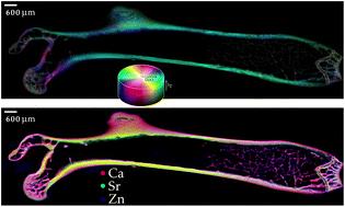
Imaging the orientations of crystallites in bone requires the usage of synchrotron X-ray radiation, which is a limited resource for researchers. Thus scans have historically been limited to either small regions or few samples. In the present study, we scan 16 full frontal cross sections of mouse femora. This makes it possible to study structure, orientation, and composition, statistically across many different bones and animals, while preserving the structural context. From the following analysis, we can deduce that while the trabecular bone in the shaft has a larger fraction of oriented crystallites than other regions in the bone, the oriented fraction is more well aligned in the cortical bone in the shaft compared to other regions in the bone. We also see that the crystallites in the cortical and trabecular bone are longer than those in the femoral head and the condyle. The study also shows a larger Sr content in the cortical bone compared to other regions, and a larger Zn content in the femoral head compared to other regions of the bones. This study shows the need for and possibility of scanning larger regions to understand bioinorganic materials.

 求助内容:
求助内容: 应助结果提醒方式:


