Lukasz Fulawka, Beata Dawiec, Wojciech Homola, Agnieszka Halon
{"title":"病例报告:富破骨巨细胞宫颈鳞状细胞癌——临床沉默的早期发现角化亚型的首例报道,并进行了详细的文献比较。","authors":"Lukasz Fulawka, Beata Dawiec, Wojciech Homola, Agnieszka Halon","doi":"10.3389/pore.2025.1612076","DOIUrl":null,"url":null,"abstract":"<p><strong>Introduction: </strong>We report the first case of an asymptomatic woman with osteoclast-like giant cell-rich cervical squamous cell carcinoma (OGC-rich cervical SCC), where the detection of cancer was made possible only by routine cytological screening. The presence of OGCs in cervical SCCs is an extremely rare phenomenon, with only 8 cases reported to date.</p><p><strong>Case description: </strong>Two consecutive liquid-based cytology revealed high-grade squamous intraepithelial lesion (HSIL). Molecular testing detected HPV 18. Colposcopic findings strongly supported the clinical diagnosis of HSIL/suspicious for invasion. Histopathological examination of biopsy samples revealed typical keratinizing-type cervical SCC morphology. The patient subsequently underwent LEEP (loop electrosurgical excision procedure). Microscopic examination of resection specimen confirmed the previous diagnosis. Moreover, groups of large multinucleated cells were observed at the periphery of some invasive nests. Most of them presented the morphology of osteoclasts, whereas some giant cells were similar to Langhans cells. All the giant cells were positive for vimentin and CD68, negative for pancytokeratin. Owing to positive margins following the LEEP procedure, the patient underwent hysterectomy via the Wertheim technique. No adjuvant treatment was applied, and after the 9-month follow-up, the patient was alive with no recurrence.</p><p><strong>Conclusion: </strong>Detailed literature review revealed that our case is the first case of keratinizing-subtype cervical OGC-rich SCC. Moreover, it is the youngest (33 yo.) patient with a significantly smaller diameter than previously reported cases. Unfortunately, owing to the small number of reported cases, the analysis did not allow us to draw conclusions about the potential prognostic or predictive value of OGC-rich morphology.</p>","PeriodicalId":19981,"journal":{"name":"Pathology & Oncology Research","volume":"31 ","pages":"1612076"},"PeriodicalIF":2.3000,"publicationDate":"2025-05-26","publicationTypes":"Journal Article","fieldsOfStudy":null,"isOpenAccess":false,"openAccessPdf":"https://www.ncbi.nlm.nih.gov/pmc/articles/PMC12146208/pdf/","citationCount":"0","resultStr":"{\"title\":\"Case Report: Osteoclastic giant cell-rich cervical squamous cell carcinoma-the first reported case of a clinically silent early-detected keratinizing subtype with a detailed literature comparison.\",\"authors\":\"Lukasz Fulawka, Beata Dawiec, Wojciech Homola, Agnieszka Halon\",\"doi\":\"10.3389/pore.2025.1612076\",\"DOIUrl\":null,\"url\":null,\"abstract\":\"<p><strong>Introduction: </strong>We report the first case of an asymptomatic woman with osteoclast-like giant cell-rich cervical squamous cell carcinoma (OGC-rich cervical SCC), where the detection of cancer was made possible only by routine cytological screening. The presence of OGCs in cervical SCCs is an extremely rare phenomenon, with only 8 cases reported to date.</p><p><strong>Case description: </strong>Two consecutive liquid-based cytology revealed high-grade squamous intraepithelial lesion (HSIL). Molecular testing detected HPV 18. Colposcopic findings strongly supported the clinical diagnosis of HSIL/suspicious for invasion. Histopathological examination of biopsy samples revealed typical keratinizing-type cervical SCC morphology. The patient subsequently underwent LEEP (loop electrosurgical excision procedure). Microscopic examination of resection specimen confirmed the previous diagnosis. Moreover, groups of large multinucleated cells were observed at the periphery of some invasive nests. Most of them presented the morphology of osteoclasts, whereas some giant cells were similar to Langhans cells. All the giant cells were positive for vimentin and CD68, negative for pancytokeratin. Owing to positive margins following the LEEP procedure, the patient underwent hysterectomy via the Wertheim technique. No adjuvant treatment was applied, and after the 9-month follow-up, the patient was alive with no recurrence.</p><p><strong>Conclusion: </strong>Detailed literature review revealed that our case is the first case of keratinizing-subtype cervical OGC-rich SCC. Moreover, it is the youngest (33 yo.) patient with a significantly smaller diameter than previously reported cases. Unfortunately, owing to the small number of reported cases, the analysis did not allow us to draw conclusions about the potential prognostic or predictive value of OGC-rich morphology.</p>\",\"PeriodicalId\":19981,\"journal\":{\"name\":\"Pathology & Oncology Research\",\"volume\":\"31 \",\"pages\":\"1612076\"},\"PeriodicalIF\":2.3000,\"publicationDate\":\"2025-05-26\",\"publicationTypes\":\"Journal Article\",\"fieldsOfStudy\":null,\"isOpenAccess\":false,\"openAccessPdf\":\"https://www.ncbi.nlm.nih.gov/pmc/articles/PMC12146208/pdf/\",\"citationCount\":\"0\",\"resultStr\":null,\"platform\":\"Semanticscholar\",\"paperid\":null,\"PeriodicalName\":\"Pathology & Oncology Research\",\"FirstCategoryId\":\"3\",\"ListUrlMain\":\"https://doi.org/10.3389/pore.2025.1612076\",\"RegionNum\":4,\"RegionCategory\":\"医学\",\"ArticlePicture\":[],\"TitleCN\":null,\"AbstractTextCN\":null,\"PMCID\":null,\"EPubDate\":\"2025/1/1 0:00:00\",\"PubModel\":\"eCollection\",\"JCR\":\"Q3\",\"JCRName\":\"ONCOLOGY\",\"Score\":null,\"Total\":0}","platform":"Semanticscholar","paperid":null,"PeriodicalName":"Pathology & Oncology Research","FirstCategoryId":"3","ListUrlMain":"https://doi.org/10.3389/pore.2025.1612076","RegionNum":4,"RegionCategory":"医学","ArticlePicture":[],"TitleCN":null,"AbstractTextCN":null,"PMCID":null,"EPubDate":"2025/1/1 0:00:00","PubModel":"eCollection","JCR":"Q3","JCRName":"ONCOLOGY","Score":null,"Total":0}
Case Report: Osteoclastic giant cell-rich cervical squamous cell carcinoma-the first reported case of a clinically silent early-detected keratinizing subtype with a detailed literature comparison.
Introduction: We report the first case of an asymptomatic woman with osteoclast-like giant cell-rich cervical squamous cell carcinoma (OGC-rich cervical SCC), where the detection of cancer was made possible only by routine cytological screening. The presence of OGCs in cervical SCCs is an extremely rare phenomenon, with only 8 cases reported to date.
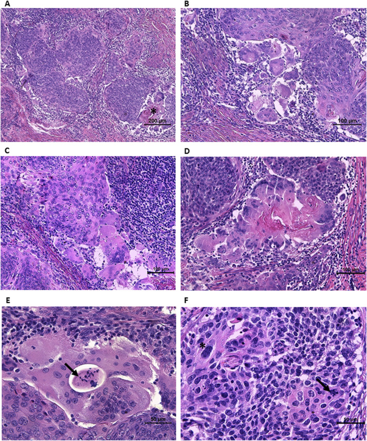
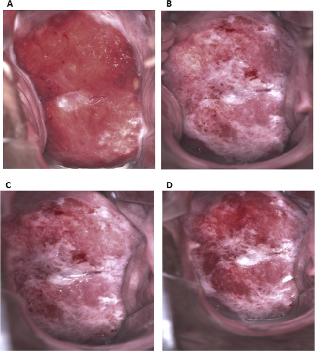
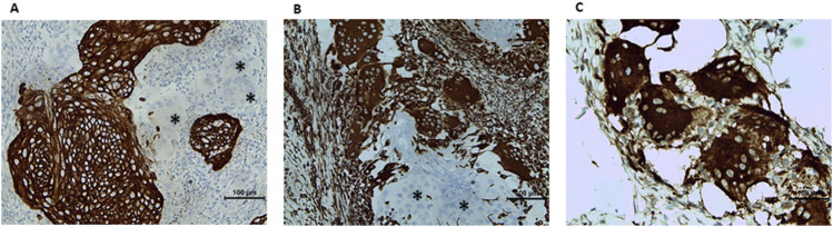
Case description: Two consecutive liquid-based cytology revealed high-grade squamous intraepithelial lesion (HSIL). Molecular testing detected HPV 18. Colposcopic findings strongly supported the clinical diagnosis of HSIL/suspicious for invasion. Histopathological examination of biopsy samples revealed typical keratinizing-type cervical SCC morphology. The patient subsequently underwent LEEP (loop electrosurgical excision procedure). Microscopic examination of resection specimen confirmed the previous diagnosis. Moreover, groups of large multinucleated cells were observed at the periphery of some invasive nests. Most of them presented the morphology of osteoclasts, whereas some giant cells were similar to Langhans cells. All the giant cells were positive for vimentin and CD68, negative for pancytokeratin. Owing to positive margins following the LEEP procedure, the patient underwent hysterectomy via the Wertheim technique. No adjuvant treatment was applied, and after the 9-month follow-up, the patient was alive with no recurrence.
Conclusion: Detailed literature review revealed that our case is the first case of keratinizing-subtype cervical OGC-rich SCC. Moreover, it is the youngest (33 yo.) patient with a significantly smaller diameter than previously reported cases. Unfortunately, owing to the small number of reported cases, the analysis did not allow us to draw conclusions about the potential prognostic or predictive value of OGC-rich morphology.
期刊介绍:
Pathology & Oncology Research (POR) is an interdisciplinary Journal at the interface of pathology and oncology including the preclinical and translational research, diagnostics and therapy. Furthermore, POR is an international forum for the rapid communication of reviews, original research, critical and topical reports with excellence and novelty. Published quarterly, POR is dedicated to keeping scientists informed of developments on the selected biomedical fields bridging the gap between basic research and clinical medicine. It is a special aim for POR to promote pathological and oncological publishing activity of colleagues in the Central and East European region. The journal will be of interest to pathologists, and a broad range of experimental and clinical oncologists, and related experts. POR is supported by an acknowledged international advisory board and the Arányi Fundation for modern pathology.
 求助内容:
求助内容: 应助结果提醒方式:


