{"title":"应用弥散张量成像鉴别高级别胶质瘤和孤立性脑转移瘤。","authors":"Shreyas Reddy K, Sandeep S, Sunitha P Kumaran, Shravan Reddy K, Meghana Kancharla","doi":"10.1055/s-0044-1801782","DOIUrl":null,"url":null,"abstract":"<p><p><b>Background</b> Brain tumors, encompassing a spectrum of neoplastic disorders, significantly impact patient morbidity and mortality. Distinguishing between high-grade gliomas (HGGs) and solitary brain metastases (SBMs) is crucial for tailored clinical management. Conventional structural magnetic resonance imaging (MRI) faces challenges in this differentiation, leading to the exploration of advanced imaging modalities such as diffusion tensor imaging (DTI). <b>Materials and Methods</b> In this prospective study, 41 patients with solitary enhancing brain lesions underwent total or subtotal resection, confirmed by histopathology. Imaging involved a 3-Tesla MRI scanner, and DTI data were analyzed for metrics including mean diffusivity, fractional anisotropy (FA), axial diffusivity, radial diffusivity, as well as planar, spherical, and linear (CL) anisotropy coefficients. <b>Results</b> Peritumoral FA and CL exhibited significant differences ( <i>p</i> = 0.0217 and <i>p</i> = 0.039, respectively) between HGG and SBM. The area under the curve for peritumoral FA and CL in differentiating HGG and SBM were 0.2791 and 0.6984, respectively. No significant differences were observed in the other diffusion metrics. <b>Conclusion</b> This study contributes to understanding DTI-derived metrics for HGG and SBM differentiation. Peritumoral FA and CL show promise as potential discriminators, offering insights for enhanced clinical decision-making and treatment planning in brain tumor patients. Future research with larger cohorts and advanced diffusion imaging techniques could further refine these findings.</p>","PeriodicalId":94300,"journal":{"name":"Asian journal of neurosurgery","volume":"20 2","pages":"278-284"},"PeriodicalIF":0.0000,"publicationDate":"2025-01-13","publicationTypes":"Journal Article","fieldsOfStudy":null,"isOpenAccess":false,"openAccessPdf":"https://www.ncbi.nlm.nih.gov/pmc/articles/PMC12136958/pdf/","citationCount":"0","resultStr":"{\"title\":\"Utilizing Diffusion Tensor Imaging to Differentiate High-Grade Gliomas and Solitary Brain Metastases.\",\"authors\":\"Shreyas Reddy K, Sandeep S, Sunitha P Kumaran, Shravan Reddy K, Meghana Kancharla\",\"doi\":\"10.1055/s-0044-1801782\",\"DOIUrl\":null,\"url\":null,\"abstract\":\"<p><p><b>Background</b> Brain tumors, encompassing a spectrum of neoplastic disorders, significantly impact patient morbidity and mortality. Distinguishing between high-grade gliomas (HGGs) and solitary brain metastases (SBMs) is crucial for tailored clinical management. Conventional structural magnetic resonance imaging (MRI) faces challenges in this differentiation, leading to the exploration of advanced imaging modalities such as diffusion tensor imaging (DTI). <b>Materials and Methods</b> In this prospective study, 41 patients with solitary enhancing brain lesions underwent total or subtotal resection, confirmed by histopathology. Imaging involved a 3-Tesla MRI scanner, and DTI data were analyzed for metrics including mean diffusivity, fractional anisotropy (FA), axial diffusivity, radial diffusivity, as well as planar, spherical, and linear (CL) anisotropy coefficients. <b>Results</b> Peritumoral FA and CL exhibited significant differences ( <i>p</i> = 0.0217 and <i>p</i> = 0.039, respectively) between HGG and SBM. The area under the curve for peritumoral FA and CL in differentiating HGG and SBM were 0.2791 and 0.6984, respectively. No significant differences were observed in the other diffusion metrics. <b>Conclusion</b> This study contributes to understanding DTI-derived metrics for HGG and SBM differentiation. Peritumoral FA and CL show promise as potential discriminators, offering insights for enhanced clinical decision-making and treatment planning in brain tumor patients. Future research with larger cohorts and advanced diffusion imaging techniques could further refine these findings.</p>\",\"PeriodicalId\":94300,\"journal\":{\"name\":\"Asian journal of neurosurgery\",\"volume\":\"20 2\",\"pages\":\"278-284\"},\"PeriodicalIF\":0.0000,\"publicationDate\":\"2025-01-13\",\"publicationTypes\":\"Journal Article\",\"fieldsOfStudy\":null,\"isOpenAccess\":false,\"openAccessPdf\":\"https://www.ncbi.nlm.nih.gov/pmc/articles/PMC12136958/pdf/\",\"citationCount\":\"0\",\"resultStr\":null,\"platform\":\"Semanticscholar\",\"paperid\":null,\"PeriodicalName\":\"Asian journal of neurosurgery\",\"FirstCategoryId\":\"1085\",\"ListUrlMain\":\"https://doi.org/10.1055/s-0044-1801782\",\"RegionNum\":0,\"RegionCategory\":null,\"ArticlePicture\":[],\"TitleCN\":null,\"AbstractTextCN\":null,\"PMCID\":null,\"EPubDate\":\"2025/6/1 0:00:00\",\"PubModel\":\"eCollection\",\"JCR\":\"\",\"JCRName\":\"\",\"Score\":null,\"Total\":0}","platform":"Semanticscholar","paperid":null,"PeriodicalName":"Asian journal of neurosurgery","FirstCategoryId":"1085","ListUrlMain":"https://doi.org/10.1055/s-0044-1801782","RegionNum":0,"RegionCategory":null,"ArticlePicture":[],"TitleCN":null,"AbstractTextCN":null,"PMCID":null,"EPubDate":"2025/6/1 0:00:00","PubModel":"eCollection","JCR":"","JCRName":"","Score":null,"Total":0}
Utilizing Diffusion Tensor Imaging to Differentiate High-Grade Gliomas and Solitary Brain Metastases.
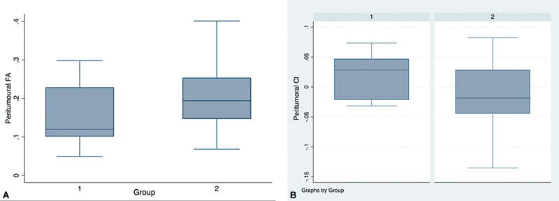
Background Brain tumors, encompassing a spectrum of neoplastic disorders, significantly impact patient morbidity and mortality. Distinguishing between high-grade gliomas (HGGs) and solitary brain metastases (SBMs) is crucial for tailored clinical management. Conventional structural magnetic resonance imaging (MRI) faces challenges in this differentiation, leading to the exploration of advanced imaging modalities such as diffusion tensor imaging (DTI). Materials and Methods In this prospective study, 41 patients with solitary enhancing brain lesions underwent total or subtotal resection, confirmed by histopathology. Imaging involved a 3-Tesla MRI scanner, and DTI data were analyzed for metrics including mean diffusivity, fractional anisotropy (FA), axial diffusivity, radial diffusivity, as well as planar, spherical, and linear (CL) anisotropy coefficients. Results Peritumoral FA and CL exhibited significant differences ( p = 0.0217 and p = 0.039, respectively) between HGG and SBM. The area under the curve for peritumoral FA and CL in differentiating HGG and SBM were 0.2791 and 0.6984, respectively. No significant differences were observed in the other diffusion metrics. Conclusion This study contributes to understanding DTI-derived metrics for HGG and SBM differentiation. Peritumoral FA and CL show promise as potential discriminators, offering insights for enhanced clinical decision-making and treatment planning in brain tumor patients. Future research with larger cohorts and advanced diffusion imaging techniques could further refine these findings.

 求助内容:
求助内容: 应助结果提醒方式:


