{"title":"术中超声在Lhermitte-Duclos病外科治疗中的应用1例报告。","authors":"Parth Parikh, Naren Nayak, Shubham Goyal, Neelanjana Ghosh, Rahul Pandey","doi":"10.1055/s-0045-1805088","DOIUrl":null,"url":null,"abstract":"<p><p>Lhermitte-Duclos disease (LDD) is a rare, autosomal dominant, dysplastic gangliocytoma of the cerebellum. It is a slow-growing benign tumor. The challenges in the surgical resection of these tumors lie in accurately delineating the tumor margin from the normal cerebellar parenchyma. Intraoperative ultrasound has the potential to overcome these limitations. A 30-year-old woman was diagnosed as having LDD showing a typical \"tigroid\" appearance on MRI. Intraoperative ultrasound was used to delineate the tumor margins and near-total resection was done via right suboccipital craniectomy. The diagnosis was confirmed on histopathological examination. Postoperatively there were no neurological deficits, and the patient is on regular follow-up for screening of Cowden's syndrome. We report this case to highlight the undervalued utility of intraoperative ultrasonography while dealing with patients with LDD. This modality serves as an effective tool to maximize the extent of resection without adding to postoperative morbidity.</p>","PeriodicalId":94300,"journal":{"name":"Asian journal of neurosurgery","volume":"20 2","pages":"413-416"},"PeriodicalIF":0.0000,"publicationDate":"2025-03-18","publicationTypes":"Journal Article","fieldsOfStudy":null,"isOpenAccess":false,"openAccessPdf":"https://www.ncbi.nlm.nih.gov/pmc/articles/PMC12136961/pdf/","citationCount":"0","resultStr":"{\"title\":\"Utility of Intraoperative Ultrasound in Surgical Management of Lhermitte-Duclos Disease: A Case Report.\",\"authors\":\"Parth Parikh, Naren Nayak, Shubham Goyal, Neelanjana Ghosh, Rahul Pandey\",\"doi\":\"10.1055/s-0045-1805088\",\"DOIUrl\":null,\"url\":null,\"abstract\":\"<p><p>Lhermitte-Duclos disease (LDD) is a rare, autosomal dominant, dysplastic gangliocytoma of the cerebellum. It is a slow-growing benign tumor. The challenges in the surgical resection of these tumors lie in accurately delineating the tumor margin from the normal cerebellar parenchyma. Intraoperative ultrasound has the potential to overcome these limitations. A 30-year-old woman was diagnosed as having LDD showing a typical \\\"tigroid\\\" appearance on MRI. Intraoperative ultrasound was used to delineate the tumor margins and near-total resection was done via right suboccipital craniectomy. The diagnosis was confirmed on histopathological examination. Postoperatively there were no neurological deficits, and the patient is on regular follow-up for screening of Cowden's syndrome. We report this case to highlight the undervalued utility of intraoperative ultrasonography while dealing with patients with LDD. This modality serves as an effective tool to maximize the extent of resection without adding to postoperative morbidity.</p>\",\"PeriodicalId\":94300,\"journal\":{\"name\":\"Asian journal of neurosurgery\",\"volume\":\"20 2\",\"pages\":\"413-416\"},\"PeriodicalIF\":0.0000,\"publicationDate\":\"2025-03-18\",\"publicationTypes\":\"Journal Article\",\"fieldsOfStudy\":null,\"isOpenAccess\":false,\"openAccessPdf\":\"https://www.ncbi.nlm.nih.gov/pmc/articles/PMC12136961/pdf/\",\"citationCount\":\"0\",\"resultStr\":null,\"platform\":\"Semanticscholar\",\"paperid\":null,\"PeriodicalName\":\"Asian journal of neurosurgery\",\"FirstCategoryId\":\"1085\",\"ListUrlMain\":\"https://doi.org/10.1055/s-0045-1805088\",\"RegionNum\":0,\"RegionCategory\":null,\"ArticlePicture\":[],\"TitleCN\":null,\"AbstractTextCN\":null,\"PMCID\":null,\"EPubDate\":\"2025/6/1 0:00:00\",\"PubModel\":\"eCollection\",\"JCR\":\"\",\"JCRName\":\"\",\"Score\":null,\"Total\":0}","platform":"Semanticscholar","paperid":null,"PeriodicalName":"Asian journal of neurosurgery","FirstCategoryId":"1085","ListUrlMain":"https://doi.org/10.1055/s-0045-1805088","RegionNum":0,"RegionCategory":null,"ArticlePicture":[],"TitleCN":null,"AbstractTextCN":null,"PMCID":null,"EPubDate":"2025/6/1 0:00:00","PubModel":"eCollection","JCR":"","JCRName":"","Score":null,"Total":0}
Utility of Intraoperative Ultrasound in Surgical Management of Lhermitte-Duclos Disease: A Case Report.
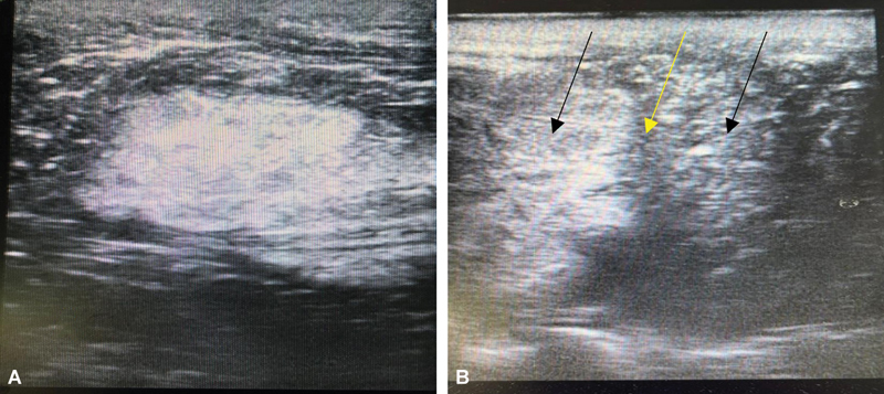
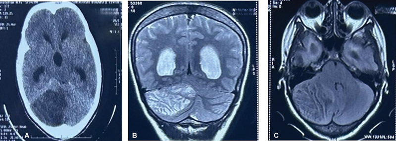
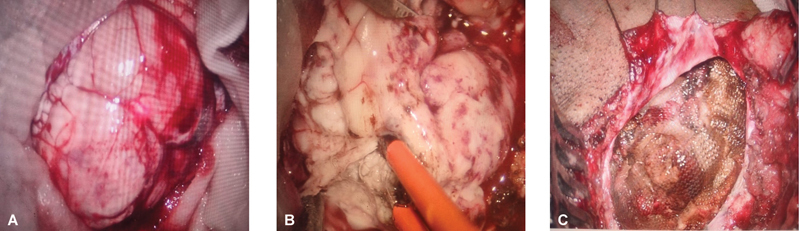
Lhermitte-Duclos disease (LDD) is a rare, autosomal dominant, dysplastic gangliocytoma of the cerebellum. It is a slow-growing benign tumor. The challenges in the surgical resection of these tumors lie in accurately delineating the tumor margin from the normal cerebellar parenchyma. Intraoperative ultrasound has the potential to overcome these limitations. A 30-year-old woman was diagnosed as having LDD showing a typical "tigroid" appearance on MRI. Intraoperative ultrasound was used to delineate the tumor margins and near-total resection was done via right suboccipital craniectomy. The diagnosis was confirmed on histopathological examination. Postoperatively there were no neurological deficits, and the patient is on regular follow-up for screening of Cowden's syndrome. We report this case to highlight the undervalued utility of intraoperative ultrasonography while dealing with patients with LDD. This modality serves as an effective tool to maximize the extent of resection without adding to postoperative morbidity.

 求助内容:
求助内容: 应助结果提醒方式:


